
癌症化学预防(专业版)
癌症化学预防在1976年由Dr. Michael Sporn创立,至今被美国国家癌症研究所及其它多个机构所公认。
英文名称:Cancer Chemoprevention
癌症化学预防包括一、二、三级预防如下:
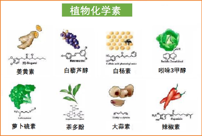
化学预防使用补充剂主要包括:植物化学素、维生素类和矿物质…等,路径如下:
有关详细内容如下:
1.抑制环氧合酶(COX-1和COX-2):
已知炎症在癌症的形成和发展中起着关键作用。人体内有许多炎症途径。环氧合酶(COX-2)是一种特殊的炎症途径,一直是肿瘤学领域的研究热点。最初,科学家们认为COX-2只是一种对炎症的诱导反应。现在推测COX-2在体内发挥生物功能,特别是在大脑、肾脏以及免疫系统中。当COX-2被促炎刺激(白细胞介素-1、生长因子、肿瘤坏死因子和内毒素)上调(有时是10到80倍)时,就会变得麻烦。当过表达时,COX-2参与各种途径,这些途径可能促进癌症(即血管生成)、细胞增殖和炎性前列腺素的产生1-3。28-30
越来越多的研究证明了COX-2与癌症之间的关系:
显然,COX-2抑制剂在癌症治疗中的作用值得肯定。一些开明的肿瘤学家在他们的抗癌方案中包括COX-2抑制剂,但数量仍然相对较少。传统NSAIDs的相关风险包括胃肠道穿孔、溃疡和出血,以及较不常见的肾和肝损伤,但对某些癌症患者的益处可能超过这些风险。
与COX-2一样,COX-1酶在某些细胞类型中也催化(介导)某些脂肪酸转化为炎症终产物。癌细胞有时会发生基因改变,导致它们表达更高水平的COX-1;在包括卵巢癌、结肠癌和头颈癌在内的几种癌症中观察到了这种情况18-20。45-47此外,用实验化合物选择性抑制COX-1已证明结肠癌细胞和卵巢癌细胞的生存能力显著降低20,21。47,48
对乳腺癌细胞的实验研究表明,同时抑制COX-1和COX-2可能对抑制癌细胞生长产生协同作用,以对抗癌细胞的生长22。49本研究的作者得出结论:“COX-1和COX-2抑制剂的联合作用及其对细胞周期的影响表明,这些药物可能成为乳腺癌的有效治疗方式。”
阿司匹林是一种非甾体抗炎药,但其作用是独特的,它选择性地抑制COX-1活性,同时调节COX-2的表达23。50这种双重作用的最终结果是通过COX-1减少有害代谢产物的产生,并降低总COX-2活性。由于COX-1和COX-2都是炎症性癌症细胞生长的驱动因素,阿司匹林是一种重要但未被充分重视的抗癌药物。
牛津大学的Peter Rothwell教授是阿司匹林作为抗癌作用研究领域的前沿人物。他和他的同事主要从事心血管医学研究,他们掌握了从八项大规模研究中收集的大量信息,这些研究考察了阿司匹林治疗对心血管健康的影响30。
他们的发现中最引人注目的是:
阿司匹林治疗与结肠癌预防之间的关联数据特别令人信服。Rothwell的团队发现,在20年的时间里,每天服用阿司匹林的患者患结肠癌的风险降低了24%,死于结肠癌的风险降低35%。在上结肠癌(升结肠和横结肠)中观察到最有效的预防益处24。51
单独的观察性研究表明,它对食道癌、胃癌、肺癌、乳腺癌和卵巢癌有预防作用25-27。52-54 一项2010年的研究表明,与不服用阿司匹林的男性相比,定期服用阿司匹林的男性患前列腺癌的风险降低了10%28。55另一项研究表明,长期服用者(5年以上)风险降低24%,每日服用者风险降低29%29。56
如何实施?
此外,一些天然COX-2抑制剂也具有一定作用,包括:白藜芦醇30(Dybkowska 2018)、维生素E(γ-和δ-生育酚)31,32(Das Gupta 2016;Abraham 2018),及生育三烯酚32-35(Abraham 2018;Jiang 2017;Husain 2017; De Silva 2016)。更多相关内容可参阅本网专文:癌症辅助疗法 >>
2.抑制癌基因Ras表达:
Ras蛋白家族在细胞生长的调控中起着核心作用。它通过整合控制细胞周期和增殖的调节信号来发挥这一基本作用。
Ras-Raf通路的缺陷可导致癌性生长。突变的Ras基因是最早发现的致癌基因之一,因为它们能够将细胞转化为癌症表型(即,由于基因表达扭曲而导致细胞明显改变)。编码Ras蛋白的三个基因(H、N或K-Ras)之一的突变与细胞增殖上调有关,估计在30-40%的人类癌症中发现。Ras突变发生率最高的是胰腺癌(80%)、结肠癌(50%)、甲状腺癌(50%),肺癌(40%)、肝癌(30%)、黑色素瘤(30%)和髓系白血病(30%)36-42。57-63
致癌基因和正常基因之间的差异可能很小。致癌基因最终产生的突变蛋白可能与健康蛋白仅相差一个氨基酸,但这种微妙的变化可以从根本上改变蛋白质的功能。
Ras-Raf通路是人类细胞从细胞表面向细胞核传递信号的途径。这些信号引导细胞分裂、分化,甚至程序性细胞死亡(凋亡)。
Ras基因通常在信号通路中充当指示细胞分裂的中继开关。为了响应从外部传递到细胞的刺激,细胞信号通路被激活。在没有刺激的情况下,Ras蛋白保持在“关闭”位置。突变的Ras蛋白基因的行为就像一个开关卡在“打开”位置,不断误导细胞,指示细胞在应该关闭周期的时候分裂43,44。64,65研究人员早就知道,注射针对氨基酸12的抗Ras抗体,导致过度增殖的逆转和突变细胞短暂改变为正常外观45。66最近,科学家利用K-Ras在几种类型的癌症中突变的高频率,开发了触发免疫系统攻击携带这种突变蛋白的细胞的疫苗。例如,2011年的一项研究发现,切除胰腺癌的患者在接种疫苗后10年存活的可能性(20%)比未接种疫苗的患者(0%)高得多46。67
为了建立诊断胰腺癌的新方法,对胰腺癌患者胰腺液中的K-Ras突变进行了检测。在87.8%(36/41)的患者中,胰液中K-Ras呈阳性47。68当结合粪便中的p53突变和CA 19-9(胰腺癌的血液标志物)时,可能比传统诊断方法更早地发现疾病48。69
对Ras突变基因活性的深入了解开辟了令人兴奋的治疗途径。研究人员发现,前体Ras基因必须经过几次生化修饰才能成为成熟、活性的基因。在这种成熟之后,Ras蛋白附着在细胞外膜的内表面,在那里它们可以与其他细胞蛋白相互作用并刺激细胞生长。
导致Ras基因成熟的事件分三步进行,最关键的是第一步,即法尼基化步骤。一种特殊的酶,法尼基蛋白转移酶(FPTase),可以加速反应。阻断Ras蛋白活性的一种策略是抑制FPTase。这种酶的抑制剂阻断Ras蛋白的成熟,并逆转突变Ras基因诱导的癌变44。65
许多天然物质影响Ras癌基因的活性。例如,柠檬烯是一种存在于柑橘类产品精油中的物质。柠檬烯已被证明是一种FPTase抑制剂。给患有癌症的动物施用高剂量的柠檬烯可阻断Ras的法尼基化,从而抑制细胞复制49,50。70,71姜黄素可抑制Ras的法尼基化,并导致表达Ras突变的乳腺癌细胞死亡51,52。72,73
日本研究人员研究了维生素E对肺癌小鼠体内K-Ras突变的影响。在用维生素E治疗之前,64%的小鼠存在K-Ras突变。在用维生素E治疗后,只有18%的小鼠表达K-Ras突变53。74维生素E降低了培养的黑色素瘤细胞中H-Ras蛋白的水平54。75匹兹堡Mercy医院进行的一项研究还表明,大蒜中天然存在的有机硫化物二烯丙基二硫,可以抑制p21 H-Ras致癌基因,对肿瘤生长有显著的抑制作用55。76
罗格斯(Rutgers)大学的研究人员调查了不同绿茶和红茶多酚抑制H-Ras致癌基因的能力。其研究小组发现,除表儿茶素(EC)外,绿茶和红茶中的所有主要多酚都对细胞生长表现出强烈的抑制作用56。77德克萨斯农工大学的研究人员还发现,鱼油可以减少大鼠结肠Ras膜的定位,减少肿瘤的形成。鉴于致癌Ras在癌症发展中的核心作用,ω-3脂肪酸调节Ras激活的发现可以解释为什么食用鱼油可以预防结肠癌57。78
他汀类药物是一类流行的降胆固醇药物。Mevacor(洛伐他汀)、Zocor(辛伐他汀)和Pravachol(普伐他汀)是他汀类药物,被证明可以抑制Ras癌基因的活性58。79他汀类药物阻断HMG-COA还原酶,从而消耗细胞中的法尼焦磷酸。这导致激活的法尼基化Ras的减少59。80
为了说明他汀类药物治疗的潜力,原发性肝癌患者接受了化疗药物5-FU或5-FU和40mg/d普伐他汀的联合治疗。81仅接受5-FU治疗的患者的中位生存期从9个月增加到5-FU联合他汀类药物普伐他汀(普伐他汀)治疗的18个月。2008年,德国研究人员研究了普伐他汀对晚期肝癌患者的影响60。81 131名患者单独接受化疗栓塞,52名患者接受化疗栓塞联合普伐他汀(20-40mg)。在长达五年的观察期内,单独接受化疗栓塞治疗的患者中有23.7%存活下来,而化疗栓塞和普伐他汀组的存活率为36.5%。仅化疗栓塞组的中位生存期为12个月,而普伐他汀组的中位生存期为20.9个月。
众所周知,他汀类药物会消耗辅酶Q10(CoQ10)水平,因此服用他汀类药物的人应该补充辅酶Q1061,62。有关详细说明,请参阅本网有关专文癌症辅助疗法的辅酶Q10部分。
如何实施?
可请医生开出这三种他汀类药物之一来抑制Ras癌基因活性:洛伐他丁(Mevacor),辛伐他汀(Zocor)和普伐他汀(Pravachol)。
注意:他汀类药物可能产生不良副作用。建议医生监督并仔细监测每月的血液测试(至少在最初),以评估肝功能、肌酶和血脂水平。
除了他汀类药物治疗外,还可以考虑补充以下营养素,以进一步抑制Ras癌基因的表达:
3.抑制血管生成:
血管生成—新生血管的生长,在胎儿发育过程中至关重要,但在健康成年人中发生率最低。例外情况发生在伤口愈合、炎症、心肌梗死后、女性生殖器官以及癌症等病理条件下63,64。110,111
血管生成是健康成人体内一个严格控制的过程,由内源性血管生成促进因子和抑制因子调节。癌症血管生成理论之父Judah Folkman博士表示,“血管生长是由对立因素的平衡控制的。刺激因子对抑制因子的偏爱可能是触发杠杆并开始肿瘤血管生成过程的原因。”65112
实体瘤不可能长到针头那么大而不诱导新血管的形成以满足肿瘤的营养需求66。113由于血管快速形成和肿瘤生长似乎同时发生,因此阻断新血管的生成对克服恶性肿瘤至关重要67。114
肿瘤血管生成是一系列分子和细胞事件的结果,通常由血管生成生长因子的释放引起。在癌症生长的关键阶段,信号分子从癌细胞分泌到附近的内皮细胞,以激活新血管的生长。这些血管生成生长因子向先前存在的血管方向扩散,促进新血管生长的形成68,69。115,116 VEGF和碱性成纤维细胞生长因子在许多肿瘤中表达,似乎对血管生成特别重要70。117
许多天然物质,如姜黄素、绿茶、N-乙酰半胱氨酸(NAC)、白藜芦醇、葡萄籽皮提取物和维生素D,具有抗血管生成特性。了解更多详细内容,可参阅本网有关专文:癌症辅助疗法 >>
美国FDA已经批准了一种名为Avastin(贝伐单抗)的抗血管生成药物,但它显示出严重的副作用,通常只有一般疗效。其他几种药物抑制血管生成是次要机制,有时用于癌症治疗。其中包括索拉非尼、舒尼替尼、帕唑帕尼和依维莫司。这些选择应该与医疗保健专业人员讨论,因为这些药物可能会造成相当大的副作用,并且只有FDA批准用于特定类型的癌症。
如何实施?
一些天然营养素已显示出潜在的抗血管生成作用:
4.抑制5脂氧合酶(5-LOX):
如上述关于抑制COX-2酶所讨论的,科学文献表明炎症在癌症的形成和发展中起着关键作用。
5-脂氧合酶(5-LOX)是另一种炎症酶,可促进癌症的形成和发展。花生四烯酸是一种在肉类和乳制品中高浓度存在的多不饱和脂肪,可促进5-LOX酶的升高。越来越多的研究表明,5-LOX通过几种明确的机制直接刺激前列腺癌症细胞增殖71-79。118-126此外,花生四烯酸被5-LOX代谢为5-HETE,这是前列腺癌细胞用来逃避破坏的一种有效生存因子74,80-83。121,127-130
为了对花生四烯酸过载的反应,身体会增加5-LOX等酶的产生,以降解花生四烯酸。5-LOX不仅直接刺激癌症细胞增殖74,84-93,121,131-140,而且5-LOX从花生四烯酸(如白三烯B4、5-HETE和羟基化脂肪酸)产生的分解产物会导致组织破坏、慢性炎症和肿瘤细胞对凋亡(程序性细胞破坏)的抵抗力增强73,80,94-98。120,127,141-145
研究表明,在前列腺癌高发地区,富含花生四烯酸的食物消费量最大72,73,78,84,119,120,125,131科学家试图确定5-LOX酶在前列腺癌和良性前列腺组织中的含量。研究人员使用前列腺活检样本发现,与良性组织相比,恶性前列腺组织中的5-LOX水平高出惊人的6倍。这项研究还发现,与良性前列腺组织相比,恶性前列腺组织中的5-HETE水平高2.2倍76。123科学家的结论是,5-LOX的选择性抑制剂可能有助于预防或治疗前列腺癌患者。
随着越来越多的证据表明摄入饱和脂肪会增加前列腺癌症风险,科学家们正在评估5-LOX对参与癌症细胞进展、血管生成和转移的各种生长因子的影响。一项研究发现,体内产生的表皮生长因子(EGF)等癌细胞增殖因子刺激前列腺癌细胞生长需要5-LOX活性。当5-LOX水平降低时,EGF等其他生长因子对癌症细胞的刺激作用减弱73。120
在一项小鼠研究中,5-LOX的增加导致血管内皮生长因子(VEGF)的相应增加,VEGF是肿瘤细胞用来刺激新血管形成(血管生成)进入肿瘤的关键生长因子。5-LOX抑制剂与许多其他生长因子一起可减少肿瘤血管生成99。146慢性炎症与诱导异常血管生成密切相关,癌细胞利用异常血管生成促进新血管生长(血管生成)进入肿瘤100。147
在雄激素依赖性和雄激素非依赖性人类前列腺癌细胞系中,5-LOX的抑制一直被证明可诱导快速和大量的细胞凋亡(癌细胞破坏)72,84,101-104。119,131,148-151
随着人类年龄的增长,慢性炎症过程会导致体内5-LOX的过度表达。过量的5-LOX可能导致老年男性前列腺癌的发生和发展105。152
根据5-LOX可以促进前列腺癌细胞侵袭和转移的累积知识,采取积极措施抑制这种致命酶似乎是有利的。减少体内5-LOX活性的一个关键方法是减少饱和脂肪和ω-6脂肪的消耗,这些脂肪含有高浓度的花生四烯酸和有助于花生四烯酸形成的高糖碳水化合物。另一种有价值的方法是补充鱼油,这会降低体内的5-LOX活性106,107。153,154研究表明,番茄红素和锯棕榈提取物也有助于抑制5-LOX101,108-121。148,155-168这些营养素对5-LOX的抑制可能部分解释了它们对前列腺的有利作用。
乳香(Boswellia)植物的特定提取物选择性抑制5-LOX122,123。169,170在几项控制良好的人体研究中,乳香已被证明能有效缓解各种慢性炎症疾病76,124-132。123,171-179科学家发现,乳香中负责抑制5-LOX的特定成分是AKBA(3-O-乙酰基-11-酮基-B-乳香酸)。AKBA直接与5-LOX结合并抑制其活性。其他乳香酸仅部分和不完全抑制5-LOX123,133。170,180
一种专利的乳香制剂(AprèsFlex)已经被开发出来并进入市场。它含有超过20%的AKBA,且生物利用度比标准化乳香提取物高52%134,135。181,182从而提供了更大的机会来抑制致命的5-LOX和花生四烯酸的其他促癌副产物。
如何实施?
减少摄入含有高浓度花生四烯酸的饱和脂肪和ω-6脂肪,如肉类、乳制品和蛋黄,以及高血糖指数碳水化合物。
建议补充以下营养素以抑制5-LOX酶活性:
5.抑制癌细胞转移:
手术切除原发性肿瘤一直是绝大多数癌症治疗的基石。这种方法的基本原理很简单:如果可以通过简单地将癌症从体内清除,那么就很可能治愈癌症。不幸的是,这种方法没有考虑到癌症在手术后会经常转移(扩散到不同的器官)。很多时候,转移性复发往往比原发肿瘤严重得多。事实上,对于许多癌症来说,最终致命的是转移性复发,而不是原发性肿瘤136。183
手术增加转移风险的一种机制是通过增强癌细胞粘附137。184脱离原发肿瘤的癌细胞利用粘附来增强其在远处器官形成转移的能力。这些癌症细胞必须能够聚集在一起,形成可以扩展和生长的聚落。单个癌细胞不太可能形成转移性肿瘤,就像一个人不太可能构成一个繁荣的社区一样。癌细胞使用粘附分子,如半乳糖凝集素-3,以促进它们聚集在一起的能力。这些分子存在于癌细胞表面,通过使单独的癌细胞相互粘附,起到像魔术贴一样的作用138。185
血液中循环的癌细胞(CTC)也利用半乳糖凝集素-3粘附在血管内壁上139。186 CTC粘附在血管壁上是转移过程的重要步骤。无法粘附在血管壁上的癌细胞将继续在血流中徘徊,无法形成转移。由于无法固定在血管壁上,这些循环的肿瘤细胞变得像“没有港口的船”,无法停靠。最终,血液中循环的白细胞会瞄准并摧毁CTC。如果CTC成功地与血管壁结合并穿过基底膜,它们将利用半乳糖凝集素-3粘附分子粘附到器官上,形成新的转移性癌症138。185
遗憾的是,研究表明,癌症手术会增加肿瘤细胞粘附140。187因此,对接受癌症手术的人来说,采取措施来帮助中和手术引起的癌细胞粘附的增加是至关重要的。
幸运的是,一种名为改性柑橘果胶(MCP)的天然化合物可以做到这一点。作为一种膳食纤维,柑橘果胶不能从肠道中吸收。然而,MCP已经被改变,从而可以被吸收到血液中并发挥其抗癌作用。MCP抑制癌细胞粘附的机制是通过与癌细胞表面的半乳糖凝集素-3粘附分子结合,从而防止癌症细胞粘附在一起并形成簇。MCP还可以抑制循环肿瘤细胞附着在血管内壁上。一项实验证明了这一点,MCP阻断了半乳糖凝集素-3与血管内壁的粘附,达到了惊人的95%。MCP还能显著降低乳腺癌细胞对血管壁的粘附141。188
在动物研究中取得这些令人兴奋的发现后,MCP随后在患有前列腺癌的男性中进行了测试。在这项试验中,10名患有复发性前列腺癌症的男性接受了MCP(每天14.4g)。一年后,前列腺癌进展显著改善,这是由前列腺特异性抗原(PSA)水平升高的速率降低决定的142。189随后进行了一项研究,对49名患有不同类型癌症的男性进行为期四周的MCP治疗。MCP治疗两个周期后,22%的男性病情稳定或生活质量改善;12%的患者病情稳定超过24周。该研究的作者得出结论:“MCP(改性柑橘果胶)似乎有积极的影响,特别是对晚期实体瘤患者的临床效益和生活质量。”143190
除了MCP,一种众所周知的OTC药物也可以在减少癌症细胞粘附方面发挥关键作用。西咪替丁(Tagamet)是一种历史上用于缓解胃灼热的药物。越来越多的科学证据表明,西咪替丁也具有强大的抗癌活性。
西咪替丁通过阻断血管内皮细胞表面粘附分子E-选择素(E-selectin)的表达来抑制癌细胞粘附。癌细胞粘附在E-选择素上,以粘附在血管内壁上144。191通过阻止E-选择素的表达,西咪替丁显著限制了癌细胞粘附在血管壁上的能力。这种效果类似于将维可牢搭扣(Velcro)从血管壁上移除,它通常会使循环肿瘤细胞结合。
2002年发表在《英国癌症》(British Journal of Cancer)上的一篇报告清楚地显示了西咪替丁的有效抗癌作用。在这项研究中,64名结肠癌患者接受了为期一年的西咪替丁(每天800mg)化疗或不化疗。西咪替丁组的10年生存率几乎为90%。这与对照组形成了鲜明对比,对照组的10年生存率仅为49.8%。值得注意的是,对于那些患有更具侵袭性结肠癌症的患者,西咪替丁治疗的患者的10年存活率为85%,而对照组的存活率为23%145,192“总之,这些结果表明了西咪替丁对结直肠癌症患者有益作用的潜在机制,可能是通过阻断血管内皮细胞上E-选择素的表达和抑制癌细胞的粘附。”这些发现得到了另一项针对结直肠癌患者的研究的支持,其中在手术时仅给予西咪替丁7天,三年生存率从59%提高到93%146。193
另一个导致癌症转移的主要因素是免疫功能障碍;主要是在外科手术(如切除原发性肿瘤)后立即发生的147。194具体而言,外科手术抑制自然杀伤(NK)细胞的特异性免疫细胞的数量,自然杀伤细胞是一种白细胞,其任务是寻找和摧毁癌症细胞。
为了说明NK细胞活性在对抗癌症中的重要性,发表在《乳腺癌研究与治疗》(Breast Cancer Research and Treatment)杂志上的一项研究,在乳腺癌手术后不久检测了女性的NK细胞活性。研究人员报告说,低水平的NK细胞活性与乳腺癌死亡风险增加有关144。191事实上,与癌症的实际阶段相比,NK细胞活性降低是更好的生存预测因素。在另一项令人担忧的研究中,结肠癌手术前NK细胞活性降低的个体在接下来的31个月内转移风险增加了350%148。195
一个突出的天然化合物,可以增加NK细胞的活性是PSK(蛋白结合多糖K),这是一种特别制备的云芝蘑菇提取物。在多项研究中,PSK已被证明能增强NK细胞活性149,150。196,197 PSK增强NK细胞活动的能力有助于解释为什么它被证明能显著提高癌症患者的生存率。例如,225名肺癌患者接受了有或无PSK的放射治疗(每天3g)。对于那些晚期3期肺癌患者,服用PSK的患者在五年后存活的人数(26%)是不服用PSK患者(8%)的三倍多。在那些患有1或2期较轻患者中,PSK的五年生存率增加了一倍多(39%对17%)151。198
在2008年的一项研究中,一组结肠癌患者被随机分为单独化疗或化疗加PSK,为期两年。接受PSK治疗组的10年生存率为82%。遗憾的是,单独接受化疗的患者10年生存率仅为51%152。199在《英国癌症杂志》报道的一项类似试验中,结肠癌患者单独接受化疗或联合PSK(每天3g)治疗两年。在具有更危险的3期结肠癌癌症的组中,PSK组的五年生存率为75%。相比之下,仅接受化疗的组的五年生存率仅为46%153。200其他研究表明,PSK也能提高乳腺癌、胃癌、食道癌和子宫癌的生存率154-156。201-203
附:甲基丙二酸(MMA)与转移
癌症的易感性和死亡率随着年龄的增长而大幅增加157,158。204,205这在一定程度上是由于在一生中积累的突变和暴露于诱变剂。然而,年龄引起的代谢变化似乎也在促进癌症细胞侵袭和转移方面发挥着重要作用。2020年发表在《自然》(Nature)杂志上的一项研究提供了证据,证明甲基丙二酸(MMA)是蛋白质和脂肪消化的代谢副产物,也是维生素B12缺乏的标志,随着年龄的增长,可以赋予癌细胞更具攻击性的特性159,160。206,207研究人员用年轻和老年健康供体的血清培养癌细胞,发现在老年供体的血清中培养的细胞倾向于经历类似于上皮-间质转化(EMT)的转变,这是癌症形成和发展的过程,并变得具有侵袭性和转移性161。208此外,老年的血清促进了对两种常见化疗药物的耐药性159。206
然后,研究人员分析了老年人和年轻人血清的代谢成分,以确定是什么导致了这种变化;他们发现,旧血清中包括MMA在内的三种化合物水平显著较高,其中只有MMA诱导癌细胞的上皮-间质样转化。MMA的升高可能是由于丙酸代谢途径中分解某些氨基酸和脂肪的酶的失调,和/或维生素B12缺乏,维生素B12是该途径中必要的辅因子。进一步的研究表明,MMA似乎上调了转录因子基因Sox4,这是EMT的“主调节因子”,在许多侵袭性癌症中过表达159,162,163。206,209,210有趣的是,MMA依赖血清中的脂质来穿透癌细胞159。206鉴于本研究的结果,对于老年人和/或癌症患者来说,采取措施降低MMA水平可能是明智的,例如保持足够的维生素B12水平,避免过量摄入蛋白质(尤其是动物来源的蛋白质),并可能降低胆固醇等血脂。然而,在得出关于这些方法在癌症和癌症预防中降低MMA的效用的确切结论之前,还需要更多的研究。
如何实施?
以下三种新化合物在抑制导致癌症转移的几种机制方面显示出有效性。在围手术期(手术前后)考虑这些化合物尤为重要,因为手术的一个已知后果是转移倾向增强。
了解癌症更多的治疗及防控方法,可参考本网站如下专文:
参考文献:
1. Sears B, Bill Lawren. The Zone: A Dietary Road Map.1995.
2. Newmark T, Paul Schulick. Beyond Aspirin: Nature's Answer to Arthritis, Cancer & Alzheimer's Disease 2000.
3. Chakraborti AK et al. Progress in COX-2 Inhibitors: A Journey So Far. Curr Med Chem. 2010;17(15):1563-93.
4. Tucker ON et al. Cyclooxygenase-2 expression is up-regulated in human pancreatic cancer. Cancer Res. 1999 Mar 1;59(5):987-90.
5. Gately S. The contributions of cyclooxygenase-2 to tumor angiogenesis. Cancer Metastasis Rev. 2000;19(1-2):19-27.
6. Reddy BS et al. Colon cancer: a role for cyclo-oxygenase-2-specific nonsteroidal anti-inflammatory drugs. Drugs Aging. 2000 May;16(5):329-34.
7. Sheehan KM et al. The relationship between cyclooxygenase-2 expression and colorectal cancer. JAMA. 1999 Oct 6;282(13):1254-7.
8. Brody JE. Aspirin linked to aid in colon cancer fight. New York Times. 1991;141(48, 812):A12.
9. Knorr J. Aspirin: the multi-purpose compound not just for headaches anymore. Life Extension Magazine 2000 Feb. 2000; Life Extension Magazine 2000 Feb6(2):50.
10. Agarwal B et al. Lovastatin augments sulindac-induced apoptosis in colon cancer cells and potentiates chemopreventive effects of sulindac. Gastroenterology. 1999 Oct;117(4):838-47.
11. Tsujii M et al. Cyclooxygenase regulates angiogenesis induced by colon cancer cells. Cell. 1998 May 29;93(5):705-16.
12. Valsecchi ME et al. Reduced risk of bone metastasis for patients with breast cancer who use COX-2 inhibitors. Clin Breast Cancer. 2009 Nov;9(4):225-30.
13. Edelman MJ et al. Eicosanoid modulation in advanced lung cancer: cyclooxygenase-2 expression is a positive predictive factor for celecoxib + chemotherapy—Cancer and Leukemia Group B Trial 30203. J Clin Oncol. 2008 Feb;26(6):848-55.
14. Pruthi RS e al. Phase II trial of celecoxib in prostate-specific antigen recurrent prostate cancer after definitive radiation therapy or radical prostatectomy. Clin Cancer Res. 2006 Apr;12(7 Pt 1):2172-7.
15. Manola J et al. Prognostic factors in metastatic melanoma: a pooled analysis of Eastern Cooperative Oncology Group trials. J Clin Oncol. 2000 Nov;18(22):3782-93.
16. Lai V et al. Results of a pilot study of the effects of celecoxib on cancer cachexia in patients with cancer of the head, neck, and gastrointestinal tract. Head Neck. 2008 Jan;30(1):67-74.
17. Harris RE. Cyclooxygenase-2 (cox-2) blockade in the chemoprevention of cancers of the colon, breast, prostate, and lung. Inflammopharmacology. 2009 Apr;17(2):55-67.
18. Erovic BM et al. Strong evidence for up-regulation of cyclooxygenase-1 in head and neck cancer. Eur J Clin Invest. 2008 Jan;38(1):61-6.
19. Khunnarong J et al. Expression of cyclooxygenase-1 in epithelial ovarian cancer: a clinicopathological study. Asian Pac J Cancer Prev. 2008 Oct-Dec;9(4):757-62.
20. Wu WK et al. Inhibition of cyclooxygenase-1 lowers proliferation and induces macroautophagy in colon cancer cells. Biochem Biophys Res Commun. 2009 Apr 24;382(1):79-84.
21. Li W et al. Effects of a cyclooxygenase-1-selective inhibitor in a mouse model of ovarian cancer, administered alone or in combination with ibuprofen, a nonselective cyclooxygenase inhibitor. Med Oncol. 2009;26(2):170-7.
22. McFadden DW et al. Additive effects of Cox-1 and Cox-2 inhibition on breast cancer in vitro. Int J Oncol. 2006 Oct;29(4):1019-23.
23. Wu KK et al. Aspirin and other cyclooxygenase inhibitors: new therapeutic insights. Semin Vasc Med. 2003 May;3(2):107-12.
24. Rothwell PM et al. Long-term effect of aspirin on colorectal cancer incidence and mortality: 20-year follow-up of five randomised trials. Lancet. 2010 Nov 20;376(9754):1741-50.
25. Sjodahl R. Nonsteroidal anti-inflammatory drugs and the gastrointestinal tract. Extent, mode, and dose dependence of anticancer effects. Am J Med. 2001 Jan 8;110(1A):66S-69S.
26. Corley DA et al. Protective association of aspirin/NSAIDs and esophageal cancer: a systematic review and meta-analysis. Gastroenterology. 2003 Jan;124(1):47-56.
27. Bosetti C et al. Aspirin and cancer risk: a summary review to 2007. Recent Results Cancer Res. 2009;181:231-51.
28. Dhillon PK et al. Long-term aspirin use and the risk of total, high-grade, regionally advanced and lethal prostate cancer in a prospective cohort of health professionals, 1988-2006. Int J Cancer. 2010 Dec 2.
29. Salinas CA et al. Use of aspirin and other nonsteroidal antiinflammatory medications in relation to prostate cancer risk. Am J Epidemiol. 2010 Sep 1;172(5):578-90.
30. Dybkowska E et al. The occurrence of resveratrol in foodstuffs and its potential for supporting cancer prevention and treatment. A review. Rocz Panstw Zakl Hig. 2018;69(1):5-14.
31. Das Gupta S et al. Tocopherols in cancer: an update. Mol Nutr Food Res. 2016;60(6):1354-1363.
32. Abraham A et al. Vitamin E and its anticancer effects. Crit Rev Food Sci Nutr. May 2018:1-23.
33. Jiang Q. Natural forms of vitamin E as effective agents for cancer prevention and therapy. Adv Nutr. 2017;8(6):850-867.
34. Husain K et al. delta-Tocotrienol, a natural form of vitamin E, inhibits pancreatic cancer stem-like cells and prevents pancreatic cancer metastasis. Oncotarget. 2017;8(19):31554-31567.
35. De Silva L et al. Tocotrienol and cancer metastasis. Biofactors. 2016;42(2):149-162.
36. Duursma AM et al. Ras interference as cancer therapy. Semin Cancer Biol. 2003 Aug;13(4):267-73.
37. Minamoto T et al. K-ras mutation: early detection in molecular diagnosis and risk assessment of colorectal, pancreas, and lung cancers--a review. Cancer Detect Prev. 2000;24(1):1-12.
38. Vachtenheim J. Occurrence of ras mutations in human lung cancer. Minireview. Neoplasma. 1997;44(3):145-9.
39. Bartram CR. Mutations in ras genes in myelocytic leukemias and myelodysplastic syndromes. Blood Cells. 1988;14(2-3):533-8.
40. Bos JL. ras oncogenes in human cancer: a review. Cancer Res. 1989 Sep;49(17):4682-9.
41. Hsieh JS et al. APC, K-ras, and p53 gene mutations in colorectal cancer patients: correlation to clinicopathologic features and postoperative surveillance. Am Surg. 2005 Apr;71(4):336-43.
42. Däbritz J et al. Follow-up study of K-ras mutations in the plasma of patients with pancreatic cancer: correlation with clinical features and carbohydrate antigen 19-9. Pancreas. 2009 Jul;38(5):534-41.
43. Gibbs JB et al. Farnesyltransferase inhibitors and anti-Ras therapy. Breast Cancer Res Treat. 1996;38(1):75-83.
44. Oliff A. Therapies of the Future: New Molecular Targets for Cancer Therapy. 1996.
45. Feramisco JR et al. Transient reversion of ras oncogene-induced cell transformation by antibodies specific for amino acid 12 of ras protein. Nature. 1985 Apr 18;314(6012):639-42.
46. Weden S et al. Long-term follow-up of patients with resected pancreatic cancer following vaccination against mutant K-ras. Int J Cancer. 2011 Mar 1;128(5):1120-8. doi: 10.1002/ijc.25449.
47. Lu X et al. [An application value of detecting K-ras and p53 gene mutation in the stool and pure pancreatic juice for diagnosis of early pancreatic cancer]. Zhonghua Yi Xue Za Zhi. 2001 Sep 10;81(17):1050-3.
48. Wu X et al. Evaluation of the diagnostic value of serum tumor markers, and fecal k-ras and p53 gene mutations for pancreatic cancer. Chin J Dig Dis. 2006;7(3):170-4.
49. Bland J. Farnesyl transferase inhibitors.2001;October 17-21, 2001.
50. Asamoto M et al. Mammary carcinomas induced in human c-Ha-ras proto-oncogene transgenic rats are estrogen-independent, but responsive to d-limonene treatment. Jpn J Cancer Res. 2002 Jan;93(1):32-5.
51. Kim Ms et al. Inhibition of invasion and induction of apoptosis by curcumin in H-ras-transformed MCF10A human breast epithelial cells. Arch Pharm Res. 2001 Aug;24(4):349-54.
52. Chen X et al. Inhibition of farnesyl protein transferase by monoterpene, curcumin derivatives and gallotannin. Anticancer Res. 1997 Jul-Aug;17(4A):2555-64.
53. Yano T et al. The inhibitory effect of vitamin E on K-ras mutation at an early stage of lung carcinogenesis in mice. Eur J Pharmacol. 1997 Mar;323(1):99-102.
54. Prasad KN, Cohrs RJ, Sharma OK. Decreased expressions of c-myc and H-ras oncogenes in vitamin E succinate induced morphologically differentiated murine B-16 melanoma cells in culture. Biochem Cell Biol. 1990 Nov;68(11):1250-5.
55. Singh A et al. Inhibition of deoxyglucose uptake in MCF-7 breast cancer cells by 2-methoxyestrone and 2-methoxyestrone-3-O-sulfamate. Mol Cell Endocrinol. 2000 Feb 25;160(1-2):61-6.
56. Chung JY et al. Inhibition of activator protein 1 activity and cell growth by purified green tea and black tea polyphenols in H-ras-transformed cells: structure-activity relationship and mechanisms involved. Cancer Res. 1999 Sep 15;59(18):4610-7.
57. Collett ED et al. n-6 and n-3 polyunsaturated fatty acids differentially modulate oncogenic Ras activation in colonocytes. Am J Physiol Cell Physiol. 2001 May;280(5):C1066-C1075.
58. Wang IK et al. Suppression of invasion and MMP-9 expression in NIH 3T3 and v-H-Ras 3T3 fibroblasts by lovastatin through inhibition of ras isoprenylation. Oncology. 2000 Sep;59(3):245-54.
59. Hohl RJ et al. Differential effects of monoterpenes and lovastatin on RAS processing. J Biol Chem. 1995 Jul 21;270(29):17508-12.
60. Kawata S et al. Effect of pravastatin on survival in patients with advanced hepatocellular carcinoma. A randomized controlled trial. Br J Cancer. 2001 Apr 6;84(7):886-91.
61. Skarlovnik A et al. Coenzyme Q10 supplementation decreases statin-related mild-to-moderate muscle symptoms: a randomized clinical study. Med Sci Monit. 2014;20:2183-2188.
62. Toth S et al. Addition of omega-3 fatty acid and coenzyme Q10 to statin therapy in patients with combined dyslipidemia. J Basic Clin Physiol Pharmacol. 2017;28(4):327-336.
63. Shammas NW et al. Acquired coronary angiogenesis after myocardial infarction. Cardiology. 1993;83(3):212-6.
64. Suh DY. Understanding angiogenesis and its clinical applications. Ann Clin Lab Sci. 2000 Jul;30(3):227-38.
65. Cooke R. Dr. Folkman's War: Angiogenesis and the Struggle to Defeat Cancer. Nature Medicine. 2001 May;7(5):525-6.
66. Folkman J. Tumor angiogenesis: therapeutic implications. N Engl J Med. 1971 Nov 18;285(21):1182-6
67. Cao Y et al. A review of Judah Folkman's remarkable achievements in biomedicine. Proc Natl Acad Sci U S A. 2008 Sep 9;105(36):13203-5.
68. Folkman J. The role of angiogenesis in tumor growth. Semin Cancer Biol. 1992b Apr;3(2):65-71.
69. Folkman J et al. Control of angiogenesis by heparin and other sulfated polysaccharides. Adv Exp Med Biol. 1992a;313:355-64.
70. NIH/NCI. Angiogenesis Inhibitors in Cancer Research. NIH/NCI Angiogenesis Inhibitors in Cancer Research 1998 Jul 7 Bethesda, MD: National Cancer Institute (Http://Www Slip Net/~Mcdavis/Database/Angio163 Htm).1998;Jul 7
71. Ghosh J. Rapid induction of apoptosis in prostate cancer cells by selenium: reversal by metabolites of arachidonate 5-lipoxygenase. Biochem Biophys Res Commun. 2004 Mar 12;315(3):24-35.
72. Moretti RM et al. Activation of the orphan nuclear receptor RORalpha counteracts the proliferative effect of fatty acids on prostate cancer cells: crucial role of 5-lipoxygenase. Int J Cancer. 2004 Oct;121(1):87-93.
73. Hassan S et al. Involvement of arachidonic acid metabolism and EGF receptor in neurotensin-induced prostate cancer PC3 cell growth. Regul Pept. 2006 Jan;133(1-3):105-14.
74. Matsuyama M et al. Expression of lipoxygenase in human prostate cancer and growth reduction by its inhibitors. Int J Oncol. 2004a Apr;24(4):821-7.
75. Kelavkar UP et al. Overexpression of 12/15-lipoxygenase, an ortholog of human 15-lipoxygenase-1, in the prostate tumors of TRAMP mice. Neoplasia. 2004 Nov;6(6):821-30.
76. Gupta S et al. Lipoxygenase-5 is overexpressed in prostate adenocarcinoma. Cancer. 2001 Feb 15;91(4):737-43
77. Kelavkar UP et al. Overexpression of 15-lipoxygenase-1 in PC-3 human prostate cancer cells increases tumorigenesis. Carcinogenesis. 2001 Nov;22(11):1765-73.
78. Ghosh J et al. Arachidonic acid stimulates prostate cancer cell growth: critical role of 5-lipoxygenase. Biochem Biophys Res Commun. 1997 Jun;235(2):418-23.
79. Gao X et al. Elevated 12-lipoxygenase mRNA expression correlates with advanced stage and poor differentiation of human prostate cancer. Urology. 1995;46(2):227-37.
80. Sundaram S et al. Expression of 5-oxoETE receptor in prostate cancer cells: critical role in survival. Biochem Biophys Res Commun. 2006 Jan 6;339(1):93-8.
81. Myers CE et al. Lipoxygenase inhibition in prostate cancer. Eur Urol. 1999;35(5-6):395-8.
82. Nakao-Hayashi J et al. Stimulatory effects of insulin and insulin-like growth factor I on migration and tube formation by vascular endothelial cells. Atherosclerosis. 1992 Feb;92(2-3):141-9.
83. Cohen P et al. Insulin-like growth factors (IGFs), IGF receptors, and IGF-binding proteins in primary cultures of prostate epithelial cells. J Clin Endocrinol Metab. 1991 Aug;73(2):401-7.
84. Ghosh J. Inhibition of arachidonate 5-lipoxygenase triggers prostate cancer cell death through rapid activation of c-Jun N-terminal kinase. Biochem Biophys Res Commun. 2003 Jul 25;307(2):342-9.
85. Jiang WG et al. Aberrant expression of 5-lipoxygenase-activating protein (5-LOXAP) has prognostic and survival significance in patients with breast cancer. Prostaglandins Leukot Essent Fatty Acids. 2006 Feb;74(2):125-34.
86. Yoshimura R et al. Relationship between lipoxygenase and human testicular cancer. Int J Mol Med. 2004 Mar;13(3):389-93.
87. Zhang L et al. Expression patterns of 5-lipoxygenase in human brain with traumatic injury and astrocytoma. Neuropathology. 2006 Apr;26(2):99-106.
88. Soumaoro LT et al. Expression of 5-Lipoxygenase in human colorectal cancer. World J Gastroenterol. 2006 Oct 21;12(39):6355-60.
89. Hayashi T et al. Inhibition of 5-lipoxygenase pathway suppresses the growth of bladder cancer cells. Int J Urol. 2006 Aug;13(8):1086-91.
90. Hoque A et al. Increased 5-lipoxygenase expression and induction of apoptosis by its inhibitors in esophageal cancer: a potential target for prevention. Carcinogenesis. 2005 Apr;26(4):785-91.
91. Hennig R et al. 5-Lipoxygenase and leukotriene B(4) receptor are expressed in human pancreatic cancers but not in pancreatic ducts in normal tissue. Am J Pathol. 2002 Aug;161(2):421-8.
92. Ding XZ et al. Lipoxygenase inhibitors abolish proliferation of human pancreatic cancer cells. Biochem Biophys Res Commun. 1999 Jul 22;261(1):218-23.
93. Matsuyama M et al. 5-Lipoxygenase inhibitors attenuate growth of human renal cell carcinoma and induce apoptosis through arachidonic acid pathway. Oncol Rep. 2005 Jul;14(1):73-9.
94. Zhi H et al. The deregulation of arachidonic acid metabolism-related genes in human esophageal squamous cell carcinoma. Int J Cancer. 2003 Sep 1;106(3):327-33.
95. Penglis PS et al. Differential regulation of prostaglandin E2 and thromboxane A2 production in human monocytes: implications for the use of cyclooxygenase inhibitors. J Immunol. 2000 Aug 1;165(3):1605-11.
96. Rubinsztajn R et al. [Urinary leukotriene E4 concentration in patients with bronchial asthma and intolerance of non-steroids anti-inflammatory drugs before and after oral aspirin challenge]. Pol Arch Med Wewn. 2003 Aug;110(2):849-854
97. Subbarao K et al. Role of leukotriene B4 receptors in the development of atherosclerosis: potential mechanisms. Arterioscler Thromb Vasc Biol. 2004 Feb;24(2):369-75.
98. Hu Y et al. 15-Lipoxygenase-1-mediated metabolism of docosahexaenoic acid is required for syndecan-1 signaling and apoptosis in prostate cancer cells. Carcinogenesis. 2013;34(1):176-82.
99. Ye YN et al. Contributory role of 5-lipoxygenase and its association with angiogenesis in the promotion of inflammation-associated colonic tumorigenesis by cigarette smoking. Toxicology. 2004 Oct 15;203(1-3):179-88.
100. Rajashekhar G et al. Continuous endothelial cell activation increases angiogenesis: evidence for the direct role of endothelium linking angiogenesis and inflammation. J Vasc Res. 2006;43(2):193-204.
101. Hazai E et al. Molecular modeling of the non-covalent binding of the dietary tomato carotenoids lycopene and lycophyll, and selected oxidative metabolites with 5-lipoxygenase. Bioorg Med Chem. 2006 Oct 15;14(20):6859-67.
102. Ghosh J et al. Inhibition of arachidonate 5-lipoxygenase triggers massive apoptosis in human prostate cancer cells. Proc Natl Acad Sci USA. 1998 Oct 27;95(22):13182-7.
103. Yang P et al. Inhibition of proliferation of PC3 cells by the branched-chain fatty acid, 12-methyltetradecanoic acid, is associated with inhibition of 5-lipoxygenase. Prostate. 2003 Jun 1;55(4):281-91.
104. Anderson KM et al. 5-Lipoxygenase inhibitors reduce PC-3 cell proliferation and initiate nonnecrotic cell death. Prostate. 1998;37(3):161-73.
105. Steinhilber D et al. 5-lipoxygenase: underappreciated role of a pro-inflammatory enzyme in tumorigenesis. Front Pharmacol. 2010;1:143. Epub 2010 Dec 24.
106. Taccone-Gallucci M et al. N-3 PUFAs reduce oxidative stress in ESRD patients on maintenance HD by inhibiting 5-lipoxygenase activity. Kidney Int. 2006 Apr;69(8):1450-4.
107. Calder PC. N-3 polyunsaturated fatty acids and inflammation: from molecular biology to the clinic. Lipids. 2003 Apr;38(4):343-52.
108. Jian L et al. Do dietary lycopene and other carotenoids protect against prostate cancer? Int J Cancer. 2005 Mar 1;113(6):1010-4.
109. Campbell JK et al. Tomato phytochemicals and prostate cancer risk. J Nutr. 2004 Dec;134(12 Suppl):3486S-92S.
110. Wertz K et al. Lycopene: modes of action to promote prostate health. Arch Biochem Biophys. 2004 Oct 1;430(1):127-34.
111. Binns CW et al. The relationship between dietary carotenoids and prostate cancer risk in Southeast Chinese men. Asia Pac J Clin Nutr. 2004;13(Suppl):S117.
112. Kim L et al. Effect of lycopene on prostate LNCaP cancer cells in culture. J Med Food. 2002;5(4):181-7.
113. Giovannucci E. A review of epidemiologic studies of tomatoes, lycopene, and prostate cancer. Exp Biol Med (Maywood.). 2002 Nov;227(10):852-9.
114. Vogt TM et al. Serum lycopene, other serum carotenoids, and risk of prostate cancer in US blacks and whites. Am J Epidemiol. 2002 Jun 1;155(11):1023-32.
115. Bosetti C et al. Fraction of prostate cancer incidence attributed to diet in Athens, Greece. Eur J Cancer Prev. 2000 Apr;9(2):119-23.
116. Blumenfeld AJ et al. Nutritional aspects of prostate cancer: a review. Can J Urol. 2000 Feb;7(1):927-35.
117. Agarwal S et al. Tomato lycopene and its role in human health and chronic diseases. CMAJ. 2000 Sep 19;163(6):739-44.
118. Gann PH et al. Lower prostate cancer risk in men with elevated plasma lycopene levels: results of a prospective analysis. Cancer Res. 1999 Mar 15;59(6):1225-30.
119. Giovannucci E et al. Intake of carotenoids and retinol in relation to risk of prostate cancer. J Natl Cancer Inst. 1995 Dec;87(23):1767-76.
120. Hill B et al. Effect of permixon on human prostate cell growth: lack of apoptotic action. Prostate. 2004 Sep 15;61(1):73-80.
121. Paubert-Braquet M et al. Effect of the lipidic lipidosterolic extract of Serenoa repens (Permixon) on the ionophore A23187-simulated production of leukotriene B4 (LTB4) from human polymorphonuclear neutrophils. Prostaglandins Leukot Essent Fatty Acids. 1997;57(3):299-304.
122. Safayhi H et al. Inhibition by boswellic acids of human leukocyte elastase. J Pharmacol Exp Ther. 1997 Apr;281(1):460-3.
123. Safayhi H et al. Mechanism of 5-lipoxygenase inhibition by acetyl-11-keto-beta-boswellic acid. Mol Pharmacol. 1995 Jun;47(6):1212-6.
124. Kimmatkar N et al. Efficacy and tolerability of Boswellia serrata extract in treatment of osteoarthritis of knee—a randomized double blind placebo controlled trial. Phytomedicine. 2003 Jan;10(1):3-7.
125. Ammon HP. Boswellic acids (components of frankincense) as the active principle in treatment of chronic inflammatory disease. Wien Med Wochenschr. 2002;152(15-16):373-8.
126. Wallace JM. Nutritional and botanical modulation of the inflammatory cascade--eicosanoids, cyclooxygenases, and lipoxygenases—as an adjunct in cancer therapy. Integr Cancer Ther. 2002 Mar;1(1):7-37.
127. Gerhardt H et al. Therapy of active Crohn disease with Boswellia serrata extract H 15. Z Gastroenterol. 2001 Jan;39(1):11-7.
128. Gupta I et al. Effects of Boswellia serrata gum resin in patients with bronchial asthma: results of a double-blind, placebo-controlled, 6-week clinical study. Eur J Med Res. 1998 Nov 17;3(11):511-4.
129. Kulkarni RR et al. Treatment of osteoarthritis with a herbomineral formulation: a double-blind, placebo-controlled, cross-over study. J Ethnopharmacol. 1991 May;33(1-2):91-5.
130. Park YS et al. Acetyl-11-keto-beta-boswellic acid (AKBA) is cytotoxic for meningioma cells and inhibits phosphorylation of the extracellular-signal regulated kinase 1 and 2. Adv Exp Med Biol. 2002;507:387-93.
131. Liu JJ et al. Keto- and acetyl-keto-boswellic acids inhibit proliferation and induce apoptosis in Hep G2 cells via a caspase-8 dependent pathway. Int J Mol Med. 2002 Oct;10(4):501-5.
132. Syrovets T et al. Acetyl-boswellic acids are novel catalytic inhibitors of human topoisomerases I and IIalpha. Mol Pharmacol. 2000 Jul;58(1):71-81.
133. Sailer ER et al. Acetyl-11-keto-beta-boswellic acid (AKBA): structure requirements for binding and 5-lipoxygenase inhibitory activity. Br J Pharmacol. 1996 Feb;117(4):615-8.
134. Sengupta K et al. Cellular and molecular mechanisms of anti-inflammatory effect of Aflapin: a novel Boswellia serrata extract. Mol Cell Biochem. 2011 Aug;354(1-2):189-97.
135. Krishnaraju AV et al. Safety and toxicological evaluation of Aflapin: a novel Boswellia-derived anti-inflammatory product. Toxicol Mech Methods. 2010;20(9):556-63.
136. Bird NC et al. Biology of colorectal liver metastases: A review. J Surg Oncol. 2006 Jul 1;94(1):68-80.
137. Dowdall JF et al. Soluble interleukin 6 receptor (sIL-6R) mediates colonic tumor cell adherence to the vascular endothelium: a mechanism for metastatic initiation? J Surg Res. 2002 Sep;107(1):1-6.
138. Raz A et al. Endogenous galactoside-binding lectins: a new class of functional tumor cell surface molecules related to metastasis. Cancer Metastasis Rev. 1987;6(3):433-52.
139. Yu LG et al. Galectin-3 interaction with Thomsen-Friedenreich disaccharide on cancer-associated MUC1 causes increased cancer cell endothelial adhesion. J Biol Chem. 2007 Jan 5;282(1):773-81. Epub 2006 Nov 7.
140. ten Kate M et al. Influence of proinflammatory cytokines on the adhesion of human colon carcinoma cells to lung microvascular endothelium. Int J Cancer. 2004 Dec 20;112(6):943-50.
141. Nangia-Makker P et al. Inhibition of human cancer cell growth and metastasis in nude mice by oral intake of modified citrus pectin. J Natl Cancer Inst. 2002 Dec 18;94(24):1854-62.
142. Guess BW et al. Modified citrus pectin (MCP) increases the prostate-specific antigen doubling time in men with prostate cancer: a phase II pilot study. Prostate Cancer Prostatic Dis. 2003;6(4):301-4.
143. Jackson CL et al. Pectin induces apoptosis in human prostate cancer cells: correlation of apoptotic function with pectin structure. Glycobiology. 2007;17(8):805-819.
144. Eichbaum C et al. Breast Cancer Cell-derived Cytokines, Macrophages and Cell Adhesion: Implications for Metastasis. Anticancer Res. 2011 Oct;31(10):3219-27.
145. Matsumoto S et al. Cimetidine increases survival of colorectal cancer patients with high levels of sialyl Lewis-X and sialyl Lewis-A epitope expression on tumour cells. Br J Cancer. 2002 Jan 21;86(2):161-7.
146. Adams WJ et al. Short-course cimetidine and survival with colorectal cancer. Lancet. 1994 Dec 24-31;344(8939-8940):1768-9.
147. Shakhar G et al. Potential prophylactic measures against postoperative immunosuppression: could they reduce recurrence rates in oncological patients? Ann Surg Oncol. 2003 Oct;10(8):972-92.
148. Koda K et al. Preoperative natural killer cell activity: correlation with distant metastases in curatively research colorectal carcinomas. Int Surg. 1997 Apr-Jun;82(2):190-3.
149. Fisher M et al. Anticancer effects and mechanisms of polysaccharide-K (PSK): implications of cancer immunotherapy. Anticancer Res. 2002 May-Jun;22(3):1737-54.
150. Garcia-Lora A et al. Protein-bound polysaccharide K and interleukin-2 regulate different nuclear transcription factors in the NKL human natural killer cell line. Cancer Immunol Immunother. 2001 Jun;50(4):191-8.
151. Hayakawa K et al. Effect of Krestin as adjuvant treatment following radical radiotherapy in non-small cell lung cancer patients. Cancer Detect Prev. 1997;21(1):71-7.
152. Sakai T et al. Immunochemotherapy with PSK and fluoropyrimidines improves long-term prognosis for curatively resected colorectal cancer. Cancer Biother Radiopharm. 2008 Aug;23(4):461-7.
153. Ohwada S et al. Adjuvant immunochemotherapy with oral Tegafur/Uracil plus PSK in patients with stage II or III colorectal cancer: a randomised controlled study. Br J Cancer. 2004 Mar 8;90(5):1003-10.
154. Okazaki A et al. [The effects of PS-K on long-term survival of uterine cervical cancer patients treated with radiation]. Gan No Rinsho. 1986 Feb;32(2):181-5.
155. Nakazato H et al. Efficacy of immunochemotherapy as adjuvant treatment after curative resection of gastric cancer. Study Group of Immunochemotherapy with PSK for Gastric Cancer. Lancet. 1994 May 7;343(8906):1122-6.
156. Toi M et al. Randomized adjuvant trial to evaluate the addition of tamoxifen and PSK to chemotherapy in patients with primary breast cancer. 5-Year results from the Nishi-Nippon Group of the Adjuvant Chemoendocrine Therapy for Breast Cancer Organization. Cancer. 1992 Nov 15;70(10):2475-83.
157. White MC et al. Age and cancer risk: a potentially modifiable relationship. American journal of preventive medicine. 2014;46(3 Suppl 1):S7-S15.
158. Harding C et al. Peak and decline in cancer incidence, mortality, and prevalence at old ages. Cancer. 2012;118(5):1371-1386.
159. Gomes AP et al. Age-induced accumulation of methylmalonic acid promotes tumour progression. Nature. 2020.
160. Vashi P et al. Methylmalonic Acid and Homocysteine as Indicators of Vitamin B-12 Deficiency in Cancer. PloS one. 2016;11(1):e0147843-e0147843.
161. Ye X et al. Epithelial-Mesenchymal Plasticity: A Central Regulator of Cancer Progression. Trends in cell biology. 2015;25(11):675-686.
162. Wang L et al. SOX4 is associated with poor prognosis in prostate cancer and promotes epithelial–mesenchymal transition in vitro. Prostate cancer and prostatic diseases. 2013;16(4):301-307.
163. Zhang J et al. SOX4 induces epithelial-mesenchymal transition and contributes to breast cancer progression. Cancer Res. 2012;72(17):4597-4608.
美国国立癌症研究所
www.cancer.gov
美国国立补充整体医学中心
https://nccih.nih.gov/
美国癌症学会
http://www.cancer.org
加拿大癌症学会
http://www.cancer.ca
免责声明和安全信息
定义
癌症化学预防在1976年由Dr. Michael Sporn创立,至今被美国国家癌症研究所及其它多个机构所公认。其意指利用天然、合成或生物物质来阻止、减缓或者逆转癌症发生、发展过程,从而降低癌症发生和死亡率的方法和策略。虽然癌症早期诊断、早期发现技术越来越好,手术、化疗和放疗在临床上已经应用了几十年,但不可忽视的是大多数临床中晚期患者的整体生存率仍无明显改善。因此,对癌症过程预防干预受到重视,并获得了越来越多的疗效。了解癌症化学预防
肿瘤发生是一个复杂的过程,是环境和遗传因素(占比5%以下)相互作用的结果,而环境因素是可预防和干预的。恶性肿瘤涉及致癌物质暴露和激活,DNA复合物的形成,炎症,氧化应激,基因突变和表观遗传的改变等。癌症具有鲜明的生物学特征,包括:持续的增生信号、生长抑制逃逸、细胞凋亡抵抗、永生细胞扩增、血管生成、癌细胞侵袭或转移以及免疫逃逸等。化学预防应针对所有的致癌过程和癌症发展的这些固有特征。癌症化学预防包括一、二、三级预防如下:
- 一级预防: 又称病因预防,针对风险增加、但无癌症病患史的、健康人群的主动预防。减少可能致癌的因素,从良好的生活习惯开始,包括坚持健康饮食,适当运动锻炼,控制体重,做好压力管理,适量饮酒或不饮酒,以及戒烟或避免二手烟等。一级预防的目的就是要把癌症的发生消灭在起始之前的未萌生状态。从分子、细胞水平而言,病因预防主要措施包括:
- 抗氧化、淬灭活性氧/亲电性物质,防止DNA损伤和突变,减少DNA加成物的形成。
- 增强DNA修复,提高DNA修复能力,在细胞增殖以前使损伤的DNA得以恢复正常。
- 支持致癌物代谢解毒,抑制致癌物的代谢活化或提高机体解毒酶系活性,以减少活性致癌物的生成。
- 二级预防:又称为临床前预防,主要针对极高风险人群或存在癌前病变的采取早筛查早治疗的措施,控制癌前病变、减少中晚期癌症发生。由于从正常细胞发展到危及生命的恶性肿瘤,大多经历“癌前病变(Premalignant lesion)”阶段。而从“癌前病变”发展成“侵袭性癌(Invasive carcinoma)”一般需要10年或更长的时间。“癌前病变”的一个重要特征是具有可逆性;“癌前病变”的转归可以作为肿瘤化学预防的重要标志,在癌症预防中有重要价值。从分子、细胞水平而言,临床前预防措施包括:
- 抗炎,抑制环氧合酶、脂氧合酶等
- 抗氧化,淬灭活性氧
- 抑制细胞增殖
- 诱导细胞凋亡
- 促进癌细胞分化
- 增强免疫功能
- 抑制血管生成
- 三级预防:又称为临床预防,主要适应癌症治疗后的康复,减轻疼痛等症状、防止复发和改善生存质量等。包括各种对症治疗、提高免疫力,防止恶病质、癌痛治疗等。从分子、细胞水平而言,临床前预防措施包括:
- 抑制癌症转移
- 防止耐药发生
- 降低化疗毒性
- 防止癌症复发
此外,三级预防仍然需要采用二级预防的多种措施,包括抗炎、抑制血管生成和增强免疫功能等。
化学预防路径和措施
所谓癌症的化学预防是指利用天然、合成或生物物质来阻止、减缓或者逆转癌症发生发展过程,从而降低癌症发生率和死亡率的方法策略。它是癌症研究的新的领域。化学预防使用补充剂主要包括:植物化学素、维生素类和矿物质…等,路径如下:
- 抑制环氧合酶(COX-1和COX-2);
- 抑制癌基因Ras表达;
- 抑制血管生成;
- 抑制5脂氧合酶(5-LOX);
- 抑制癌细胞转移。
有关详细内容如下:
1.抑制环氧合酶(COX-1和COX-2):
已知炎症在癌症的形成和发展中起着关键作用。人体内有许多炎症途径。环氧合酶(COX-2)是一种特殊的炎症途径,一直是肿瘤学领域的研究热点。最初,科学家们认为COX-2只是一种对炎症的诱导反应。现在推测COX-2在体内发挥生物功能,特别是在大脑、肾脏以及免疫系统中。当COX-2被促炎刺激(白细胞介素-1、生长因子、肿瘤坏死因子和内毒素)上调(有时是10到80倍)时,就会变得麻烦。当过表达时,COX-2参与各种途径,这些途径可能促进癌症(即血管生成)、细胞增殖和炎性前列腺素的产生1-3。28-30
越来越多的研究证明了COX-2与癌症之间的关系:
- 《癌症研究》(Cancer Reserch)杂志上的一篇文章表明,胰腺癌细胞中的COX-2水平是邻近正常组织的60倍4。31
- 实体瘤含有缺氧或缺氧区域(组织的氧气供应减少到低于生理水平)。缺氧促进COX-2和血管生成的上调,并建立对电离辐射的抗性5。32
- 在非甾体抗炎药(NSAIDs)类别中,有一个子类被称为COX-2抑制剂。COX-2抑制剂被广泛用于止痛,但现在已经在肿瘤学中找到了一席之地。这始于科学家们认识到,经常服用NSAIDS的人患结肠癌的风险降低了50%6。33
- 《美国医学会》(JAMA)报道,一项为期9.4年的流行病学研究表明,COX-2上调与结直肠癌患者更晚期的肿瘤分期、肿瘤大小、淋巴结转移以及生存率降低有关7。34更经常地使用阿司匹林(一种COX-2抑制剂),死于该疾病的风险降低8,9。35,36《胃肠病学》(Gastroenterology)报道的发现令人鼓舞,显示三种不同的结肠癌细胞系在缺乏COX-2时发生细胞凋亡(细胞死亡);当将洛伐他汀添加到COX-2抑制剂中时,致死率又增加了五倍10。37观察到的COX-2抑制剂的益处不仅限于结肠保护,还包括心血管系统,它们有助于维持内皮细胞功能11。38
- 2009年发表的一项突破性研究表明,接受COX-2抑制剂治疗的乳腺癌患者骨转移风险大大降低。在这项研究中,记录了未使用COX-2抑制剂治疗的乳癌患者以及在诊断为乳癌后接受COX-2抑制剂至少六个月的患者的骨转移发生率。这一发现令人震惊,接受COX-2抑制剂治疗的患者发生骨转移的可能性比未接受COX-2抑制物治疗的患者低90%12。39
- 134名晚期肺癌患者接受了单独化疗或与西乐葆(一种COX-2抑制剂)联合化疗。对于那些COX-2表达量增加的癌症患者,西乐葆治疗显著延长了生存期13。40
- 西乐葆减缓了复发性前列腺癌患者的癌症进展14,15。41,42
- 西乐葆可防止头颈癌患者的体重减轻,并改善其生活质量16。43
- 定期服用非处方OTC的NSAIDs可显著降低结肠癌风险43%,乳腺癌风险25%,肺癌风险28%,前列腺癌风险27%。此外,在一系列病例对照研究中,每天使用选择性COX-2抑制剂(塞来昔布或罗非昔布),可显著降低每种恶性肿瘤的风险。有证据表明,对COX-2具有选择性或非选择性活性的抗炎药,在结肠癌、乳腺癌、前列腺癌和肺癌的化学预防方面具有强大的潜力。一些选择性COX-2抑制剂对心血管系统构成风险的观察结果证实了COX-2阻断对癌症预防有效17。44
显然,COX-2抑制剂在癌症治疗中的作用值得肯定。一些开明的肿瘤学家在他们的抗癌方案中包括COX-2抑制剂,但数量仍然相对较少。传统NSAIDs的相关风险包括胃肠道穿孔、溃疡和出血,以及较不常见的肾和肝损伤,但对某些癌症患者的益处可能超过这些风险。
与COX-2一样,COX-1酶在某些细胞类型中也催化(介导)某些脂肪酸转化为炎症终产物。癌细胞有时会发生基因改变,导致它们表达更高水平的COX-1;在包括卵巢癌、结肠癌和头颈癌在内的几种癌症中观察到了这种情况18-20。45-47此外,用实验化合物选择性抑制COX-1已证明结肠癌细胞和卵巢癌细胞的生存能力显著降低20,21。47,48
对乳腺癌细胞的实验研究表明,同时抑制COX-1和COX-2可能对抑制癌细胞生长产生协同作用,以对抗癌细胞的生长22。49本研究的作者得出结论:“COX-1和COX-2抑制剂的联合作用及其对细胞周期的影响表明,这些药物可能成为乳腺癌的有效治疗方式。”
阿司匹林是一种非甾体抗炎药,但其作用是独特的,它选择性地抑制COX-1活性,同时调节COX-2的表达23。50这种双重作用的最终结果是通过COX-1减少有害代谢产物的产生,并降低总COX-2活性。由于COX-1和COX-2都是炎症性癌症细胞生长的驱动因素,阿司匹林是一种重要但未被充分重视的抗癌药物。
牛津大学的Peter Rothwell教授是阿司匹林作为抗癌作用研究领域的前沿人物。他和他的同事主要从事心血管医学研究,他们掌握了从八项大规模研究中收集的大量信息,这些研究考察了阿司匹林治疗对心血管健康的影响30。
他们的发现中最引人注目的是:
- 阿司匹林可将癌症死亡的总体风险降低约20%。
- 大部分益处是由于每天摄入阿司匹林五年后死亡率减少了30-40%。
- 在可获得这一时期数据的研究中,实体癌死亡率的下降保持了20年。
- 这些影响在所有研究人群中都是一致的,尽管他们的健康史各不相同。
- 每天仅75mg剂量就能产生保护作用,更高的剂量并不能增加益处。
- 癌症死亡率的减少随着年龄的增长而增加:在55-64岁的人群中观察到峰值效应,在65岁或65岁以上的人群中仍然很高。
- 阿司匹林对降低致命癌症风险的作用非常强大,足以显著降低各种原因造成的死亡率。
阿司匹林治疗与结肠癌预防之间的关联数据特别令人信服。Rothwell的团队发现,在20年的时间里,每天服用阿司匹林的患者患结肠癌的风险降低了24%,死于结肠癌的风险降低35%。在上结肠癌(升结肠和横结肠)中观察到最有效的预防益处24。51
单独的观察性研究表明,它对食道癌、胃癌、肺癌、乳腺癌和卵巢癌有预防作用25-27。52-54 一项2010年的研究表明,与不服用阿司匹林的男性相比,定期服用阿司匹林的男性患前列腺癌的风险降低了10%28。55另一项研究表明,长期服用者(5年以上)风险降低24%,每日服用者风险降低29%29。56
如何实施?
- 每天服用小剂量阿司匹林,并且,
- 请医生开出COX-2抑制药物之一:(1).依托度酸(Lodine XL,罗丁), 1000mg,每日一次,或(2).西乐葆,每12小时100-200mg
此外,一些天然COX-2抑制剂也具有一定作用,包括:白藜芦醇30(Dybkowska 2018)、维生素E(γ-和δ-生育酚)31,32(Das Gupta 2016;Abraham 2018),及生育三烯酚32-35(Abraham 2018;Jiang 2017;Husain 2017; De Silva 2016)。更多相关内容可参阅本网专文:癌症辅助疗法 >>
2.抑制癌基因Ras表达:
Ras蛋白家族在细胞生长的调控中起着核心作用。它通过整合控制细胞周期和增殖的调节信号来发挥这一基本作用。
Ras-Raf通路的缺陷可导致癌性生长。突变的Ras基因是最早发现的致癌基因之一,因为它们能够将细胞转化为癌症表型(即,由于基因表达扭曲而导致细胞明显改变)。编码Ras蛋白的三个基因(H、N或K-Ras)之一的突变与细胞增殖上调有关,估计在30-40%的人类癌症中发现。Ras突变发生率最高的是胰腺癌(80%)、结肠癌(50%)、甲状腺癌(50%),肺癌(40%)、肝癌(30%)、黑色素瘤(30%)和髓系白血病(30%)36-42。57-63
致癌基因和正常基因之间的差异可能很小。致癌基因最终产生的突变蛋白可能与健康蛋白仅相差一个氨基酸,但这种微妙的变化可以从根本上改变蛋白质的功能。
Ras-Raf通路是人类细胞从细胞表面向细胞核传递信号的途径。这些信号引导细胞分裂、分化,甚至程序性细胞死亡(凋亡)。
Ras基因通常在信号通路中充当指示细胞分裂的中继开关。为了响应从外部传递到细胞的刺激,细胞信号通路被激活。在没有刺激的情况下,Ras蛋白保持在“关闭”位置。突变的Ras蛋白基因的行为就像一个开关卡在“打开”位置,不断误导细胞,指示细胞在应该关闭周期的时候分裂43,44。64,65研究人员早就知道,注射针对氨基酸12的抗Ras抗体,导致过度增殖的逆转和突变细胞短暂改变为正常外观45。66最近,科学家利用K-Ras在几种类型的癌症中突变的高频率,开发了触发免疫系统攻击携带这种突变蛋白的细胞的疫苗。例如,2011年的一项研究发现,切除胰腺癌的患者在接种疫苗后10年存活的可能性(20%)比未接种疫苗的患者(0%)高得多46。67
为了建立诊断胰腺癌的新方法,对胰腺癌患者胰腺液中的K-Ras突变进行了检测。在87.8%(36/41)的患者中,胰液中K-Ras呈阳性47。68当结合粪便中的p53突变和CA 19-9(胰腺癌的血液标志物)时,可能比传统诊断方法更早地发现疾病48。69
对Ras突变基因活性的深入了解开辟了令人兴奋的治疗途径。研究人员发现,前体Ras基因必须经过几次生化修饰才能成为成熟、活性的基因。在这种成熟之后,Ras蛋白附着在细胞外膜的内表面,在那里它们可以与其他细胞蛋白相互作用并刺激细胞生长。
导致Ras基因成熟的事件分三步进行,最关键的是第一步,即法尼基化步骤。一种特殊的酶,法尼基蛋白转移酶(FPTase),可以加速反应。阻断Ras蛋白活性的一种策略是抑制FPTase。这种酶的抑制剂阻断Ras蛋白的成熟,并逆转突变Ras基因诱导的癌变44。65
许多天然物质影响Ras癌基因的活性。例如,柠檬烯是一种存在于柑橘类产品精油中的物质。柠檬烯已被证明是一种FPTase抑制剂。给患有癌症的动物施用高剂量的柠檬烯可阻断Ras的法尼基化,从而抑制细胞复制49,50。70,71姜黄素可抑制Ras的法尼基化,并导致表达Ras突变的乳腺癌细胞死亡51,52。72,73
日本研究人员研究了维生素E对肺癌小鼠体内K-Ras突变的影响。在用维生素E治疗之前,64%的小鼠存在K-Ras突变。在用维生素E治疗后,只有18%的小鼠表达K-Ras突变53。74维生素E降低了培养的黑色素瘤细胞中H-Ras蛋白的水平54。75匹兹堡Mercy医院进行的一项研究还表明,大蒜中天然存在的有机硫化物二烯丙基二硫,可以抑制p21 H-Ras致癌基因,对肿瘤生长有显著的抑制作用55。76
罗格斯(Rutgers)大学的研究人员调查了不同绿茶和红茶多酚抑制H-Ras致癌基因的能力。其研究小组发现,除表儿茶素(EC)外,绿茶和红茶中的所有主要多酚都对细胞生长表现出强烈的抑制作用56。77德克萨斯农工大学的研究人员还发现,鱼油可以减少大鼠结肠Ras膜的定位,减少肿瘤的形成。鉴于致癌Ras在癌症发展中的核心作用,ω-3脂肪酸调节Ras激活的发现可以解释为什么食用鱼油可以预防结肠癌57。78
他汀类药物是一类流行的降胆固醇药物。Mevacor(洛伐他汀)、Zocor(辛伐他汀)和Pravachol(普伐他汀)是他汀类药物,被证明可以抑制Ras癌基因的活性58。79他汀类药物阻断HMG-COA还原酶,从而消耗细胞中的法尼焦磷酸。这导致激活的法尼基化Ras的减少59。80
为了说明他汀类药物治疗的潜力,原发性肝癌患者接受了化疗药物5-FU或5-FU和40mg/d普伐他汀的联合治疗。81仅接受5-FU治疗的患者的中位生存期从9个月增加到5-FU联合他汀类药物普伐他汀(普伐他汀)治疗的18个月。2008年,德国研究人员研究了普伐他汀对晚期肝癌患者的影响60。81 131名患者单独接受化疗栓塞,52名患者接受化疗栓塞联合普伐他汀(20-40mg)。在长达五年的观察期内,单独接受化疗栓塞治疗的患者中有23.7%存活下来,而化疗栓塞和普伐他汀组的存活率为36.5%。仅化疗栓塞组的中位生存期为12个月,而普伐他汀组的中位生存期为20.9个月。
众所周知,他汀类药物会消耗辅酶Q10(CoQ10)水平,因此服用他汀类药物的人应该补充辅酶Q1061,62。有关详细说明,请参阅本网有关专文癌症辅助疗法的辅酶Q10部分。
如何实施?
可请医生开出这三种他汀类药物之一来抑制Ras癌基因活性:洛伐他丁(Mevacor),辛伐他汀(Zocor)和普伐他汀(Pravachol)。
注意:他汀类药物可能产生不良副作用。建议医生监督并仔细监测每月的血液测试(至少在最初),以评估肝功能、肌酶和血脂水平。
除了他汀类药物治疗外,还可以考虑补充以下营养素,以进一步抑制Ras癌基因的表达:
- 姜黄素:400–800mg/天
- 鱼油:每天2100mg EPA和1500mg DHA,随餐服用
- 绿茶(标提物):每天725–1450mg (EGCG)
- 大蒜(陈化蒜提取物):每天2400mg,随餐食用
- 维生素E(混合型):每天400–1000IU,含天然α-生育酚,以及至少200mg γ-生育酚,随餐服用。
3.抑制血管生成:
血管生成—新生血管的生长,在胎儿发育过程中至关重要,但在健康成年人中发生率最低。例外情况发生在伤口愈合、炎症、心肌梗死后、女性生殖器官以及癌症等病理条件下63,64。110,111
血管生成是健康成人体内一个严格控制的过程,由内源性血管生成促进因子和抑制因子调节。癌症血管生成理论之父Judah Folkman博士表示,“血管生长是由对立因素的平衡控制的。刺激因子对抑制因子的偏爱可能是触发杠杆并开始肿瘤血管生成过程的原因。”65112
实体瘤不可能长到针头那么大而不诱导新血管的形成以满足肿瘤的营养需求66。113由于血管快速形成和肿瘤生长似乎同时发生,因此阻断新血管的生成对克服恶性肿瘤至关重要67。114
肿瘤血管生成是一系列分子和细胞事件的结果,通常由血管生成生长因子的释放引起。在癌症生长的关键阶段,信号分子从癌细胞分泌到附近的内皮细胞,以激活新血管的生长。这些血管生成生长因子向先前存在的血管方向扩散,促进新血管生长的形成68,69。115,116 VEGF和碱性成纤维细胞生长因子在许多肿瘤中表达,似乎对血管生成特别重要70。117
许多天然物质,如姜黄素、绿茶、N-乙酰半胱氨酸(NAC)、白藜芦醇、葡萄籽皮提取物和维生素D,具有抗血管生成特性。了解更多详细内容,可参阅本网有关专文:癌症辅助疗法 >>
美国FDA已经批准了一种名为Avastin(贝伐单抗)的抗血管生成药物,但它显示出严重的副作用,通常只有一般疗效。其他几种药物抑制血管生成是次要机制,有时用于癌症治疗。其中包括索拉非尼、舒尼替尼、帕唑帕尼和依维莫司。这些选择应该与医疗保健专业人员讨论,因为这些药物可能会造成相当大的副作用,并且只有FDA批准用于特定类型的癌症。
如何实施?
一些天然营养素已显示出潜在的抗血管生成作用:
- 绿茶(标提物):725–1450mg/天
- 姜黄素:400–800mg/天
- 维生素D:5000–8000IU/天,取决于血液水平
- 葡萄籽提取物(种子和果皮):150–300mg/天
- N-乙酰半胱氨酸:600–1200mg/天
4.抑制5脂氧合酶(5-LOX):
如上述关于抑制COX-2酶所讨论的,科学文献表明炎症在癌症的形成和发展中起着关键作用。
5-脂氧合酶(5-LOX)是另一种炎症酶,可促进癌症的形成和发展。花生四烯酸是一种在肉类和乳制品中高浓度存在的多不饱和脂肪,可促进5-LOX酶的升高。越来越多的研究表明,5-LOX通过几种明确的机制直接刺激前列腺癌症细胞增殖71-79。118-126此外,花生四烯酸被5-LOX代谢为5-HETE,这是前列腺癌细胞用来逃避破坏的一种有效生存因子74,80-83。121,127-130
为了对花生四烯酸过载的反应,身体会增加5-LOX等酶的产生,以降解花生四烯酸。5-LOX不仅直接刺激癌症细胞增殖74,84-93,121,131-140,而且5-LOX从花生四烯酸(如白三烯B4、5-HETE和羟基化脂肪酸)产生的分解产物会导致组织破坏、慢性炎症和肿瘤细胞对凋亡(程序性细胞破坏)的抵抗力增强73,80,94-98。120,127,141-145
研究表明,在前列腺癌高发地区,富含花生四烯酸的食物消费量最大72,73,78,84,119,120,125,131科学家试图确定5-LOX酶在前列腺癌和良性前列腺组织中的含量。研究人员使用前列腺活检样本发现,与良性组织相比,恶性前列腺组织中的5-LOX水平高出惊人的6倍。这项研究还发现,与良性前列腺组织相比,恶性前列腺组织中的5-HETE水平高2.2倍76。123科学家的结论是,5-LOX的选择性抑制剂可能有助于预防或治疗前列腺癌患者。
随着越来越多的证据表明摄入饱和脂肪会增加前列腺癌症风险,科学家们正在评估5-LOX对参与癌症细胞进展、血管生成和转移的各种生长因子的影响。一项研究发现,体内产生的表皮生长因子(EGF)等癌细胞增殖因子刺激前列腺癌细胞生长需要5-LOX活性。当5-LOX水平降低时,EGF等其他生长因子对癌症细胞的刺激作用减弱73。120
在一项小鼠研究中,5-LOX的增加导致血管内皮生长因子(VEGF)的相应增加,VEGF是肿瘤细胞用来刺激新血管形成(血管生成)进入肿瘤的关键生长因子。5-LOX抑制剂与许多其他生长因子一起可减少肿瘤血管生成99。146慢性炎症与诱导异常血管生成密切相关,癌细胞利用异常血管生成促进新血管生长(血管生成)进入肿瘤100。147
在雄激素依赖性和雄激素非依赖性人类前列腺癌细胞系中,5-LOX的抑制一直被证明可诱导快速和大量的细胞凋亡(癌细胞破坏)72,84,101-104。119,131,148-151
随着人类年龄的增长,慢性炎症过程会导致体内5-LOX的过度表达。过量的5-LOX可能导致老年男性前列腺癌的发生和发展105。152
根据5-LOX可以促进前列腺癌细胞侵袭和转移的累积知识,采取积极措施抑制这种致命酶似乎是有利的。减少体内5-LOX活性的一个关键方法是减少饱和脂肪和ω-6脂肪的消耗,这些脂肪含有高浓度的花生四烯酸和有助于花生四烯酸形成的高糖碳水化合物。另一种有价值的方法是补充鱼油,这会降低体内的5-LOX活性106,107。153,154研究表明,番茄红素和锯棕榈提取物也有助于抑制5-LOX101,108-121。148,155-168这些营养素对5-LOX的抑制可能部分解释了它们对前列腺的有利作用。
乳香(Boswellia)植物的特定提取物选择性抑制5-LOX122,123。169,170在几项控制良好的人体研究中,乳香已被证明能有效缓解各种慢性炎症疾病76,124-132。123,171-179科学家发现,乳香中负责抑制5-LOX的特定成分是AKBA(3-O-乙酰基-11-酮基-B-乳香酸)。AKBA直接与5-LOX结合并抑制其活性。其他乳香酸仅部分和不完全抑制5-LOX123,133。170,180
一种专利的乳香制剂(AprèsFlex)已经被开发出来并进入市场。它含有超过20%的AKBA,且生物利用度比标准化乳香提取物高52%134,135。181,182从而提供了更大的机会来抑制致命的5-LOX和花生四烯酸的其他促癌副产物。
如何实施?
减少摄入含有高浓度花生四烯酸的饱和脂肪和ω-6脂肪,如肉类、乳制品和蛋黄,以及高血糖指数碳水化合物。
建议补充以下营养素以抑制5-LOX酶活性:
5.抑制癌细胞转移:
手术切除原发性肿瘤一直是绝大多数癌症治疗的基石。这种方法的基本原理很简单:如果可以通过简单地将癌症从体内清除,那么就很可能治愈癌症。不幸的是,这种方法没有考虑到癌症在手术后会经常转移(扩散到不同的器官)。很多时候,转移性复发往往比原发肿瘤严重得多。事实上,对于许多癌症来说,最终致命的是转移性复发,而不是原发性肿瘤136。183
手术增加转移风险的一种机制是通过增强癌细胞粘附137。184脱离原发肿瘤的癌细胞利用粘附来增强其在远处器官形成转移的能力。这些癌症细胞必须能够聚集在一起,形成可以扩展和生长的聚落。单个癌细胞不太可能形成转移性肿瘤,就像一个人不太可能构成一个繁荣的社区一样。癌细胞使用粘附分子,如半乳糖凝集素-3,以促进它们聚集在一起的能力。这些分子存在于癌细胞表面,通过使单独的癌细胞相互粘附,起到像魔术贴一样的作用138。185
血液中循环的癌细胞(CTC)也利用半乳糖凝集素-3粘附在血管内壁上139。186 CTC粘附在血管壁上是转移过程的重要步骤。无法粘附在血管壁上的癌细胞将继续在血流中徘徊,无法形成转移。由于无法固定在血管壁上,这些循环的肿瘤细胞变得像“没有港口的船”,无法停靠。最终,血液中循环的白细胞会瞄准并摧毁CTC。如果CTC成功地与血管壁结合并穿过基底膜,它们将利用半乳糖凝集素-3粘附分子粘附到器官上,形成新的转移性癌症138。185
遗憾的是,研究表明,癌症手术会增加肿瘤细胞粘附140。187因此,对接受癌症手术的人来说,采取措施来帮助中和手术引起的癌细胞粘附的增加是至关重要的。
幸运的是,一种名为改性柑橘果胶(MCP)的天然化合物可以做到这一点。作为一种膳食纤维,柑橘果胶不能从肠道中吸收。然而,MCP已经被改变,从而可以被吸收到血液中并发挥其抗癌作用。MCP抑制癌细胞粘附的机制是通过与癌细胞表面的半乳糖凝集素-3粘附分子结合,从而防止癌症细胞粘附在一起并形成簇。MCP还可以抑制循环肿瘤细胞附着在血管内壁上。一项实验证明了这一点,MCP阻断了半乳糖凝集素-3与血管内壁的粘附,达到了惊人的95%。MCP还能显著降低乳腺癌细胞对血管壁的粘附141。188
在动物研究中取得这些令人兴奋的发现后,MCP随后在患有前列腺癌的男性中进行了测试。在这项试验中,10名患有复发性前列腺癌症的男性接受了MCP(每天14.4g)。一年后,前列腺癌进展显著改善,这是由前列腺特异性抗原(PSA)水平升高的速率降低决定的142。189随后进行了一项研究,对49名患有不同类型癌症的男性进行为期四周的MCP治疗。MCP治疗两个周期后,22%的男性病情稳定或生活质量改善;12%的患者病情稳定超过24周。该研究的作者得出结论:“MCP(改性柑橘果胶)似乎有积极的影响,特别是对晚期实体瘤患者的临床效益和生活质量。”143190
除了MCP,一种众所周知的OTC药物也可以在减少癌症细胞粘附方面发挥关键作用。西咪替丁(Tagamet)是一种历史上用于缓解胃灼热的药物。越来越多的科学证据表明,西咪替丁也具有强大的抗癌活性。
西咪替丁通过阻断血管内皮细胞表面粘附分子E-选择素(E-selectin)的表达来抑制癌细胞粘附。癌细胞粘附在E-选择素上,以粘附在血管内壁上144。191通过阻止E-选择素的表达,西咪替丁显著限制了癌细胞粘附在血管壁上的能力。这种效果类似于将维可牢搭扣(Velcro)从血管壁上移除,它通常会使循环肿瘤细胞结合。
2002年发表在《英国癌症》(British Journal of Cancer)上的一篇报告清楚地显示了西咪替丁的有效抗癌作用。在这项研究中,64名结肠癌患者接受了为期一年的西咪替丁(每天800mg)化疗或不化疗。西咪替丁组的10年生存率几乎为90%。这与对照组形成了鲜明对比,对照组的10年生存率仅为49.8%。值得注意的是,对于那些患有更具侵袭性结肠癌症的患者,西咪替丁治疗的患者的10年存活率为85%,而对照组的存活率为23%145,192“总之,这些结果表明了西咪替丁对结直肠癌症患者有益作用的潜在机制,可能是通过阻断血管内皮细胞上E-选择素的表达和抑制癌细胞的粘附。”这些发现得到了另一项针对结直肠癌患者的研究的支持,其中在手术时仅给予西咪替丁7天,三年生存率从59%提高到93%146。193
另一个导致癌症转移的主要因素是免疫功能障碍;主要是在外科手术(如切除原发性肿瘤)后立即发生的147。194具体而言,外科手术抑制自然杀伤(NK)细胞的特异性免疫细胞的数量,自然杀伤细胞是一种白细胞,其任务是寻找和摧毁癌症细胞。
为了说明NK细胞活性在对抗癌症中的重要性,发表在《乳腺癌研究与治疗》(Breast Cancer Research and Treatment)杂志上的一项研究,在乳腺癌手术后不久检测了女性的NK细胞活性。研究人员报告说,低水平的NK细胞活性与乳腺癌死亡风险增加有关144。191事实上,与癌症的实际阶段相比,NK细胞活性降低是更好的生存预测因素。在另一项令人担忧的研究中,结肠癌手术前NK细胞活性降低的个体在接下来的31个月内转移风险增加了350%148。195
一个突出的天然化合物,可以增加NK细胞的活性是PSK(蛋白结合多糖K),这是一种特别制备的云芝蘑菇提取物。在多项研究中,PSK已被证明能增强NK细胞活性149,150。196,197 PSK增强NK细胞活动的能力有助于解释为什么它被证明能显著提高癌症患者的生存率。例如,225名肺癌患者接受了有或无PSK的放射治疗(每天3g)。对于那些晚期3期肺癌患者,服用PSK的患者在五年后存活的人数(26%)是不服用PSK患者(8%)的三倍多。在那些患有1或2期较轻患者中,PSK的五年生存率增加了一倍多(39%对17%)151。198
在2008年的一项研究中,一组结肠癌患者被随机分为单独化疗或化疗加PSK,为期两年。接受PSK治疗组的10年生存率为82%。遗憾的是,单独接受化疗的患者10年生存率仅为51%152。199在《英国癌症杂志》报道的一项类似试验中,结肠癌患者单独接受化疗或联合PSK(每天3g)治疗两年。在具有更危险的3期结肠癌癌症的组中,PSK组的五年生存率为75%。相比之下,仅接受化疗的组的五年生存率仅为46%153。200其他研究表明,PSK也能提高乳腺癌、胃癌、食道癌和子宫癌的生存率154-156。201-203
附:甲基丙二酸(MMA)与转移
癌症的易感性和死亡率随着年龄的增长而大幅增加157,158。204,205这在一定程度上是由于在一生中积累的突变和暴露于诱变剂。然而,年龄引起的代谢变化似乎也在促进癌症细胞侵袭和转移方面发挥着重要作用。2020年发表在《自然》(Nature)杂志上的一项研究提供了证据,证明甲基丙二酸(MMA)是蛋白质和脂肪消化的代谢副产物,也是维生素B12缺乏的标志,随着年龄的增长,可以赋予癌细胞更具攻击性的特性159,160。206,207研究人员用年轻和老年健康供体的血清培养癌细胞,发现在老年供体的血清中培养的细胞倾向于经历类似于上皮-间质转化(EMT)的转变,这是癌症形成和发展的过程,并变得具有侵袭性和转移性161。208此外,老年的血清促进了对两种常见化疗药物的耐药性159。206
然后,研究人员分析了老年人和年轻人血清的代谢成分,以确定是什么导致了这种变化;他们发现,旧血清中包括MMA在内的三种化合物水平显著较高,其中只有MMA诱导癌细胞的上皮-间质样转化。MMA的升高可能是由于丙酸代谢途径中分解某些氨基酸和脂肪的酶的失调,和/或维生素B12缺乏,维生素B12是该途径中必要的辅因子。进一步的研究表明,MMA似乎上调了转录因子基因Sox4,这是EMT的“主调节因子”,在许多侵袭性癌症中过表达159,162,163。206,209,210有趣的是,MMA依赖血清中的脂质来穿透癌细胞159。206鉴于本研究的结果,对于老年人和/或癌症患者来说,采取措施降低MMA水平可能是明智的,例如保持足够的维生素B12水平,避免过量摄入蛋白质(尤其是动物来源的蛋白质),并可能降低胆固醇等血脂。然而,在得出关于这些方法在癌症和癌症预防中降低MMA的效用的确切结论之前,还需要更多的研究。
如何实施?
以下三种新化合物在抑制导致癌症转移的几种机制方面显示出有效性。在围手术期(手术前后)考虑这些化合物尤为重要,因为手术的一个已知后果是转移倾向增强。
化学预防的综合干预
基于循证医学和营养学有关文献综合,可点击其综合干预方案如下:了解癌症更多的治疗及防控方法,可参考本网站如下专文:
参考文献:
1. Sears B, Bill Lawren. The Zone: A Dietary Road Map.1995.
2. Newmark T, Paul Schulick. Beyond Aspirin: Nature's Answer to Arthritis, Cancer & Alzheimer's Disease 2000.
3. Chakraborti AK et al. Progress in COX-2 Inhibitors: A Journey So Far. Curr Med Chem. 2010;17(15):1563-93.
4. Tucker ON et al. Cyclooxygenase-2 expression is up-regulated in human pancreatic cancer. Cancer Res. 1999 Mar 1;59(5):987-90.
5. Gately S. The contributions of cyclooxygenase-2 to tumor angiogenesis. Cancer Metastasis Rev. 2000;19(1-2):19-27.
6. Reddy BS et al. Colon cancer: a role for cyclo-oxygenase-2-specific nonsteroidal anti-inflammatory drugs. Drugs Aging. 2000 May;16(5):329-34.
7. Sheehan KM et al. The relationship between cyclooxygenase-2 expression and colorectal cancer. JAMA. 1999 Oct 6;282(13):1254-7.
8. Brody JE. Aspirin linked to aid in colon cancer fight. New York Times. 1991;141(48, 812):A12.
9. Knorr J. Aspirin: the multi-purpose compound not just for headaches anymore. Life Extension Magazine 2000 Feb. 2000; Life Extension Magazine 2000 Feb6(2):50.
10. Agarwal B et al. Lovastatin augments sulindac-induced apoptosis in colon cancer cells and potentiates chemopreventive effects of sulindac. Gastroenterology. 1999 Oct;117(4):838-47.
11. Tsujii M et al. Cyclooxygenase regulates angiogenesis induced by colon cancer cells. Cell. 1998 May 29;93(5):705-16.
12. Valsecchi ME et al. Reduced risk of bone metastasis for patients with breast cancer who use COX-2 inhibitors. Clin Breast Cancer. 2009 Nov;9(4):225-30.
13. Edelman MJ et al. Eicosanoid modulation in advanced lung cancer: cyclooxygenase-2 expression is a positive predictive factor for celecoxib + chemotherapy—Cancer and Leukemia Group B Trial 30203. J Clin Oncol. 2008 Feb;26(6):848-55.
14. Pruthi RS e al. Phase II trial of celecoxib in prostate-specific antigen recurrent prostate cancer after definitive radiation therapy or radical prostatectomy. Clin Cancer Res. 2006 Apr;12(7 Pt 1):2172-7.
15. Manola J et al. Prognostic factors in metastatic melanoma: a pooled analysis of Eastern Cooperative Oncology Group trials. J Clin Oncol. 2000 Nov;18(22):3782-93.
16. Lai V et al. Results of a pilot study of the effects of celecoxib on cancer cachexia in patients with cancer of the head, neck, and gastrointestinal tract. Head Neck. 2008 Jan;30(1):67-74.
17. Harris RE. Cyclooxygenase-2 (cox-2) blockade in the chemoprevention of cancers of the colon, breast, prostate, and lung. Inflammopharmacology. 2009 Apr;17(2):55-67.
18. Erovic BM et al. Strong evidence for up-regulation of cyclooxygenase-1 in head and neck cancer. Eur J Clin Invest. 2008 Jan;38(1):61-6.
19. Khunnarong J et al. Expression of cyclooxygenase-1 in epithelial ovarian cancer: a clinicopathological study. Asian Pac J Cancer Prev. 2008 Oct-Dec;9(4):757-62.
20. Wu WK et al. Inhibition of cyclooxygenase-1 lowers proliferation and induces macroautophagy in colon cancer cells. Biochem Biophys Res Commun. 2009 Apr 24;382(1):79-84.
21. Li W et al. Effects of a cyclooxygenase-1-selective inhibitor in a mouse model of ovarian cancer, administered alone or in combination with ibuprofen, a nonselective cyclooxygenase inhibitor. Med Oncol. 2009;26(2):170-7.
22. McFadden DW et al. Additive effects of Cox-1 and Cox-2 inhibition on breast cancer in vitro. Int J Oncol. 2006 Oct;29(4):1019-23.
23. Wu KK et al. Aspirin and other cyclooxygenase inhibitors: new therapeutic insights. Semin Vasc Med. 2003 May;3(2):107-12.
24. Rothwell PM et al. Long-term effect of aspirin on colorectal cancer incidence and mortality: 20-year follow-up of five randomised trials. Lancet. 2010 Nov 20;376(9754):1741-50.
25. Sjodahl R. Nonsteroidal anti-inflammatory drugs and the gastrointestinal tract. Extent, mode, and dose dependence of anticancer effects. Am J Med. 2001 Jan 8;110(1A):66S-69S.
26. Corley DA et al. Protective association of aspirin/NSAIDs and esophageal cancer: a systematic review and meta-analysis. Gastroenterology. 2003 Jan;124(1):47-56.
27. Bosetti C et al. Aspirin and cancer risk: a summary review to 2007. Recent Results Cancer Res. 2009;181:231-51.
28. Dhillon PK et al. Long-term aspirin use and the risk of total, high-grade, regionally advanced and lethal prostate cancer in a prospective cohort of health professionals, 1988-2006. Int J Cancer. 2010 Dec 2.
29. Salinas CA et al. Use of aspirin and other nonsteroidal antiinflammatory medications in relation to prostate cancer risk. Am J Epidemiol. 2010 Sep 1;172(5):578-90.
30. Dybkowska E et al. The occurrence of resveratrol in foodstuffs and its potential for supporting cancer prevention and treatment. A review. Rocz Panstw Zakl Hig. 2018;69(1):5-14.
31. Das Gupta S et al. Tocopherols in cancer: an update. Mol Nutr Food Res. 2016;60(6):1354-1363.
32. Abraham A et al. Vitamin E and its anticancer effects. Crit Rev Food Sci Nutr. May 2018:1-23.
33. Jiang Q. Natural forms of vitamin E as effective agents for cancer prevention and therapy. Adv Nutr. 2017;8(6):850-867.
34. Husain K et al. delta-Tocotrienol, a natural form of vitamin E, inhibits pancreatic cancer stem-like cells and prevents pancreatic cancer metastasis. Oncotarget. 2017;8(19):31554-31567.
35. De Silva L et al. Tocotrienol and cancer metastasis. Biofactors. 2016;42(2):149-162.
36. Duursma AM et al. Ras interference as cancer therapy. Semin Cancer Biol. 2003 Aug;13(4):267-73.
37. Minamoto T et al. K-ras mutation: early detection in molecular diagnosis and risk assessment of colorectal, pancreas, and lung cancers--a review. Cancer Detect Prev. 2000;24(1):1-12.
38. Vachtenheim J. Occurrence of ras mutations in human lung cancer. Minireview. Neoplasma. 1997;44(3):145-9.
39. Bartram CR. Mutations in ras genes in myelocytic leukemias and myelodysplastic syndromes. Blood Cells. 1988;14(2-3):533-8.
40. Bos JL. ras oncogenes in human cancer: a review. Cancer Res. 1989 Sep;49(17):4682-9.
41. Hsieh JS et al. APC, K-ras, and p53 gene mutations in colorectal cancer patients: correlation to clinicopathologic features and postoperative surveillance. Am Surg. 2005 Apr;71(4):336-43.
42. Däbritz J et al. Follow-up study of K-ras mutations in the plasma of patients with pancreatic cancer: correlation with clinical features and carbohydrate antigen 19-9. Pancreas. 2009 Jul;38(5):534-41.
43. Gibbs JB et al. Farnesyltransferase inhibitors and anti-Ras therapy. Breast Cancer Res Treat. 1996;38(1):75-83.
44. Oliff A. Therapies of the Future: New Molecular Targets for Cancer Therapy. 1996.
45. Feramisco JR et al. Transient reversion of ras oncogene-induced cell transformation by antibodies specific for amino acid 12 of ras protein. Nature. 1985 Apr 18;314(6012):639-42.
46. Weden S et al. Long-term follow-up of patients with resected pancreatic cancer following vaccination against mutant K-ras. Int J Cancer. 2011 Mar 1;128(5):1120-8. doi: 10.1002/ijc.25449.
47. Lu X et al. [An application value of detecting K-ras and p53 gene mutation in the stool and pure pancreatic juice for diagnosis of early pancreatic cancer]. Zhonghua Yi Xue Za Zhi. 2001 Sep 10;81(17):1050-3.
48. Wu X et al. Evaluation of the diagnostic value of serum tumor markers, and fecal k-ras and p53 gene mutations for pancreatic cancer. Chin J Dig Dis. 2006;7(3):170-4.
49. Bland J. Farnesyl transferase inhibitors.2001;October 17-21, 2001.
50. Asamoto M et al. Mammary carcinomas induced in human c-Ha-ras proto-oncogene transgenic rats are estrogen-independent, but responsive to d-limonene treatment. Jpn J Cancer Res. 2002 Jan;93(1):32-5.
51. Kim Ms et al. Inhibition of invasion and induction of apoptosis by curcumin in H-ras-transformed MCF10A human breast epithelial cells. Arch Pharm Res. 2001 Aug;24(4):349-54.
52. Chen X et al. Inhibition of farnesyl protein transferase by monoterpene, curcumin derivatives and gallotannin. Anticancer Res. 1997 Jul-Aug;17(4A):2555-64.
53. Yano T et al. The inhibitory effect of vitamin E on K-ras mutation at an early stage of lung carcinogenesis in mice. Eur J Pharmacol. 1997 Mar;323(1):99-102.
54. Prasad KN, Cohrs RJ, Sharma OK. Decreased expressions of c-myc and H-ras oncogenes in vitamin E succinate induced morphologically differentiated murine B-16 melanoma cells in culture. Biochem Cell Biol. 1990 Nov;68(11):1250-5.
55. Singh A et al. Inhibition of deoxyglucose uptake in MCF-7 breast cancer cells by 2-methoxyestrone and 2-methoxyestrone-3-O-sulfamate. Mol Cell Endocrinol. 2000 Feb 25;160(1-2):61-6.
56. Chung JY et al. Inhibition of activator protein 1 activity and cell growth by purified green tea and black tea polyphenols in H-ras-transformed cells: structure-activity relationship and mechanisms involved. Cancer Res. 1999 Sep 15;59(18):4610-7.
57. Collett ED et al. n-6 and n-3 polyunsaturated fatty acids differentially modulate oncogenic Ras activation in colonocytes. Am J Physiol Cell Physiol. 2001 May;280(5):C1066-C1075.
58. Wang IK et al. Suppression of invasion and MMP-9 expression in NIH 3T3 and v-H-Ras 3T3 fibroblasts by lovastatin through inhibition of ras isoprenylation. Oncology. 2000 Sep;59(3):245-54.
59. Hohl RJ et al. Differential effects of monoterpenes and lovastatin on RAS processing. J Biol Chem. 1995 Jul 21;270(29):17508-12.
60. Kawata S et al. Effect of pravastatin on survival in patients with advanced hepatocellular carcinoma. A randomized controlled trial. Br J Cancer. 2001 Apr 6;84(7):886-91.
61. Skarlovnik A et al. Coenzyme Q10 supplementation decreases statin-related mild-to-moderate muscle symptoms: a randomized clinical study. Med Sci Monit. 2014;20:2183-2188.
62. Toth S et al. Addition of omega-3 fatty acid and coenzyme Q10 to statin therapy in patients with combined dyslipidemia. J Basic Clin Physiol Pharmacol. 2017;28(4):327-336.
63. Shammas NW et al. Acquired coronary angiogenesis after myocardial infarction. Cardiology. 1993;83(3):212-6.
64. Suh DY. Understanding angiogenesis and its clinical applications. Ann Clin Lab Sci. 2000 Jul;30(3):227-38.
65. Cooke R. Dr. Folkman's War: Angiogenesis and the Struggle to Defeat Cancer. Nature Medicine. 2001 May;7(5):525-6.
66. Folkman J. Tumor angiogenesis: therapeutic implications. N Engl J Med. 1971 Nov 18;285(21):1182-6
67. Cao Y et al. A review of Judah Folkman's remarkable achievements in biomedicine. Proc Natl Acad Sci U S A. 2008 Sep 9;105(36):13203-5.
68. Folkman J. The role of angiogenesis in tumor growth. Semin Cancer Biol. 1992b Apr;3(2):65-71.
69. Folkman J et al. Control of angiogenesis by heparin and other sulfated polysaccharides. Adv Exp Med Biol. 1992a;313:355-64.
70. NIH/NCI. Angiogenesis Inhibitors in Cancer Research. NIH/NCI Angiogenesis Inhibitors in Cancer Research 1998 Jul 7 Bethesda, MD: National Cancer Institute (Http://Www Slip Net/~Mcdavis/Database/Angio163 Htm).1998;Jul 7
71. Ghosh J. Rapid induction of apoptosis in prostate cancer cells by selenium: reversal by metabolites of arachidonate 5-lipoxygenase. Biochem Biophys Res Commun. 2004 Mar 12;315(3):24-35.
72. Moretti RM et al. Activation of the orphan nuclear receptor RORalpha counteracts the proliferative effect of fatty acids on prostate cancer cells: crucial role of 5-lipoxygenase. Int J Cancer. 2004 Oct;121(1):87-93.
73. Hassan S et al. Involvement of arachidonic acid metabolism and EGF receptor in neurotensin-induced prostate cancer PC3 cell growth. Regul Pept. 2006 Jan;133(1-3):105-14.
74. Matsuyama M et al. Expression of lipoxygenase in human prostate cancer and growth reduction by its inhibitors. Int J Oncol. 2004a Apr;24(4):821-7.
75. Kelavkar UP et al. Overexpression of 12/15-lipoxygenase, an ortholog of human 15-lipoxygenase-1, in the prostate tumors of TRAMP mice. Neoplasia. 2004 Nov;6(6):821-30.
76. Gupta S et al. Lipoxygenase-5 is overexpressed in prostate adenocarcinoma. Cancer. 2001 Feb 15;91(4):737-43
77. Kelavkar UP et al. Overexpression of 15-lipoxygenase-1 in PC-3 human prostate cancer cells increases tumorigenesis. Carcinogenesis. 2001 Nov;22(11):1765-73.
78. Ghosh J et al. Arachidonic acid stimulates prostate cancer cell growth: critical role of 5-lipoxygenase. Biochem Biophys Res Commun. 1997 Jun;235(2):418-23.
79. Gao X et al. Elevated 12-lipoxygenase mRNA expression correlates with advanced stage and poor differentiation of human prostate cancer. Urology. 1995;46(2):227-37.
80. Sundaram S et al. Expression of 5-oxoETE receptor in prostate cancer cells: critical role in survival. Biochem Biophys Res Commun. 2006 Jan 6;339(1):93-8.
81. Myers CE et al. Lipoxygenase inhibition in prostate cancer. Eur Urol. 1999;35(5-6):395-8.
82. Nakao-Hayashi J et al. Stimulatory effects of insulin and insulin-like growth factor I on migration and tube formation by vascular endothelial cells. Atherosclerosis. 1992 Feb;92(2-3):141-9.
83. Cohen P et al. Insulin-like growth factors (IGFs), IGF receptors, and IGF-binding proteins in primary cultures of prostate epithelial cells. J Clin Endocrinol Metab. 1991 Aug;73(2):401-7.
84. Ghosh J. Inhibition of arachidonate 5-lipoxygenase triggers prostate cancer cell death through rapid activation of c-Jun N-terminal kinase. Biochem Biophys Res Commun. 2003 Jul 25;307(2):342-9.
85. Jiang WG et al. Aberrant expression of 5-lipoxygenase-activating protein (5-LOXAP) has prognostic and survival significance in patients with breast cancer. Prostaglandins Leukot Essent Fatty Acids. 2006 Feb;74(2):125-34.
86. Yoshimura R et al. Relationship between lipoxygenase and human testicular cancer. Int J Mol Med. 2004 Mar;13(3):389-93.
87. Zhang L et al. Expression patterns of 5-lipoxygenase in human brain with traumatic injury and astrocytoma. Neuropathology. 2006 Apr;26(2):99-106.
88. Soumaoro LT et al. Expression of 5-Lipoxygenase in human colorectal cancer. World J Gastroenterol. 2006 Oct 21;12(39):6355-60.
89. Hayashi T et al. Inhibition of 5-lipoxygenase pathway suppresses the growth of bladder cancer cells. Int J Urol. 2006 Aug;13(8):1086-91.
90. Hoque A et al. Increased 5-lipoxygenase expression and induction of apoptosis by its inhibitors in esophageal cancer: a potential target for prevention. Carcinogenesis. 2005 Apr;26(4):785-91.
91. Hennig R et al. 5-Lipoxygenase and leukotriene B(4) receptor are expressed in human pancreatic cancers but not in pancreatic ducts in normal tissue. Am J Pathol. 2002 Aug;161(2):421-8.
92. Ding XZ et al. Lipoxygenase inhibitors abolish proliferation of human pancreatic cancer cells. Biochem Biophys Res Commun. 1999 Jul 22;261(1):218-23.
93. Matsuyama M et al. 5-Lipoxygenase inhibitors attenuate growth of human renal cell carcinoma and induce apoptosis through arachidonic acid pathway. Oncol Rep. 2005 Jul;14(1):73-9.
94. Zhi H et al. The deregulation of arachidonic acid metabolism-related genes in human esophageal squamous cell carcinoma. Int J Cancer. 2003 Sep 1;106(3):327-33.
95. Penglis PS et al. Differential regulation of prostaglandin E2 and thromboxane A2 production in human monocytes: implications for the use of cyclooxygenase inhibitors. J Immunol. 2000 Aug 1;165(3):1605-11.
96. Rubinsztajn R et al. [Urinary leukotriene E4 concentration in patients with bronchial asthma and intolerance of non-steroids anti-inflammatory drugs before and after oral aspirin challenge]. Pol Arch Med Wewn. 2003 Aug;110(2):849-854
97. Subbarao K et al. Role of leukotriene B4 receptors in the development of atherosclerosis: potential mechanisms. Arterioscler Thromb Vasc Biol. 2004 Feb;24(2):369-75.
98. Hu Y et al. 15-Lipoxygenase-1-mediated metabolism of docosahexaenoic acid is required for syndecan-1 signaling and apoptosis in prostate cancer cells. Carcinogenesis. 2013;34(1):176-82.
99. Ye YN et al. Contributory role of 5-lipoxygenase and its association with angiogenesis in the promotion of inflammation-associated colonic tumorigenesis by cigarette smoking. Toxicology. 2004 Oct 15;203(1-3):179-88.
100. Rajashekhar G et al. Continuous endothelial cell activation increases angiogenesis: evidence for the direct role of endothelium linking angiogenesis and inflammation. J Vasc Res. 2006;43(2):193-204.
101. Hazai E et al. Molecular modeling of the non-covalent binding of the dietary tomato carotenoids lycopene and lycophyll, and selected oxidative metabolites with 5-lipoxygenase. Bioorg Med Chem. 2006 Oct 15;14(20):6859-67.
102. Ghosh J et al. Inhibition of arachidonate 5-lipoxygenase triggers massive apoptosis in human prostate cancer cells. Proc Natl Acad Sci USA. 1998 Oct 27;95(22):13182-7.
103. Yang P et al. Inhibition of proliferation of PC3 cells by the branched-chain fatty acid, 12-methyltetradecanoic acid, is associated with inhibition of 5-lipoxygenase. Prostate. 2003 Jun 1;55(4):281-91.
104. Anderson KM et al. 5-Lipoxygenase inhibitors reduce PC-3 cell proliferation and initiate nonnecrotic cell death. Prostate. 1998;37(3):161-73.
105. Steinhilber D et al. 5-lipoxygenase: underappreciated role of a pro-inflammatory enzyme in tumorigenesis. Front Pharmacol. 2010;1:143. Epub 2010 Dec 24.
106. Taccone-Gallucci M et al. N-3 PUFAs reduce oxidative stress in ESRD patients on maintenance HD by inhibiting 5-lipoxygenase activity. Kidney Int. 2006 Apr;69(8):1450-4.
107. Calder PC. N-3 polyunsaturated fatty acids and inflammation: from molecular biology to the clinic. Lipids. 2003 Apr;38(4):343-52.
108. Jian L et al. Do dietary lycopene and other carotenoids protect against prostate cancer? Int J Cancer. 2005 Mar 1;113(6):1010-4.
109. Campbell JK et al. Tomato phytochemicals and prostate cancer risk. J Nutr. 2004 Dec;134(12 Suppl):3486S-92S.
110. Wertz K et al. Lycopene: modes of action to promote prostate health. Arch Biochem Biophys. 2004 Oct 1;430(1):127-34.
111. Binns CW et al. The relationship between dietary carotenoids and prostate cancer risk in Southeast Chinese men. Asia Pac J Clin Nutr. 2004;13(Suppl):S117.
112. Kim L et al. Effect of lycopene on prostate LNCaP cancer cells in culture. J Med Food. 2002;5(4):181-7.
113. Giovannucci E. A review of epidemiologic studies of tomatoes, lycopene, and prostate cancer. Exp Biol Med (Maywood.). 2002 Nov;227(10):852-9.
114. Vogt TM et al. Serum lycopene, other serum carotenoids, and risk of prostate cancer in US blacks and whites. Am J Epidemiol. 2002 Jun 1;155(11):1023-32.
115. Bosetti C et al. Fraction of prostate cancer incidence attributed to diet in Athens, Greece. Eur J Cancer Prev. 2000 Apr;9(2):119-23.
116. Blumenfeld AJ et al. Nutritional aspects of prostate cancer: a review. Can J Urol. 2000 Feb;7(1):927-35.
117. Agarwal S et al. Tomato lycopene and its role in human health and chronic diseases. CMAJ. 2000 Sep 19;163(6):739-44.
118. Gann PH et al. Lower prostate cancer risk in men with elevated plasma lycopene levels: results of a prospective analysis. Cancer Res. 1999 Mar 15;59(6):1225-30.
119. Giovannucci E et al. Intake of carotenoids and retinol in relation to risk of prostate cancer. J Natl Cancer Inst. 1995 Dec;87(23):1767-76.
120. Hill B et al. Effect of permixon on human prostate cell growth: lack of apoptotic action. Prostate. 2004 Sep 15;61(1):73-80.
121. Paubert-Braquet M et al. Effect of the lipidic lipidosterolic extract of Serenoa repens (Permixon) on the ionophore A23187-simulated production of leukotriene B4 (LTB4) from human polymorphonuclear neutrophils. Prostaglandins Leukot Essent Fatty Acids. 1997;57(3):299-304.
122. Safayhi H et al. Inhibition by boswellic acids of human leukocyte elastase. J Pharmacol Exp Ther. 1997 Apr;281(1):460-3.
123. Safayhi H et al. Mechanism of 5-lipoxygenase inhibition by acetyl-11-keto-beta-boswellic acid. Mol Pharmacol. 1995 Jun;47(6):1212-6.
124. Kimmatkar N et al. Efficacy and tolerability of Boswellia serrata extract in treatment of osteoarthritis of knee—a randomized double blind placebo controlled trial. Phytomedicine. 2003 Jan;10(1):3-7.
125. Ammon HP. Boswellic acids (components of frankincense) as the active principle in treatment of chronic inflammatory disease. Wien Med Wochenschr. 2002;152(15-16):373-8.
126. Wallace JM. Nutritional and botanical modulation of the inflammatory cascade--eicosanoids, cyclooxygenases, and lipoxygenases—as an adjunct in cancer therapy. Integr Cancer Ther. 2002 Mar;1(1):7-37.
127. Gerhardt H et al. Therapy of active Crohn disease with Boswellia serrata extract H 15. Z Gastroenterol. 2001 Jan;39(1):11-7.
128. Gupta I et al. Effects of Boswellia serrata gum resin in patients with bronchial asthma: results of a double-blind, placebo-controlled, 6-week clinical study. Eur J Med Res. 1998 Nov 17;3(11):511-4.
129. Kulkarni RR et al. Treatment of osteoarthritis with a herbomineral formulation: a double-blind, placebo-controlled, cross-over study. J Ethnopharmacol. 1991 May;33(1-2):91-5.
130. Park YS et al. Acetyl-11-keto-beta-boswellic acid (AKBA) is cytotoxic for meningioma cells and inhibits phosphorylation of the extracellular-signal regulated kinase 1 and 2. Adv Exp Med Biol. 2002;507:387-93.
131. Liu JJ et al. Keto- and acetyl-keto-boswellic acids inhibit proliferation and induce apoptosis in Hep G2 cells via a caspase-8 dependent pathway. Int J Mol Med. 2002 Oct;10(4):501-5.
132. Syrovets T et al. Acetyl-boswellic acids are novel catalytic inhibitors of human topoisomerases I and IIalpha. Mol Pharmacol. 2000 Jul;58(1):71-81.
133. Sailer ER et al. Acetyl-11-keto-beta-boswellic acid (AKBA): structure requirements for binding and 5-lipoxygenase inhibitory activity. Br J Pharmacol. 1996 Feb;117(4):615-8.
134. Sengupta K et al. Cellular and molecular mechanisms of anti-inflammatory effect of Aflapin: a novel Boswellia serrata extract. Mol Cell Biochem. 2011 Aug;354(1-2):189-97.
135. Krishnaraju AV et al. Safety and toxicological evaluation of Aflapin: a novel Boswellia-derived anti-inflammatory product. Toxicol Mech Methods. 2010;20(9):556-63.
136. Bird NC et al. Biology of colorectal liver metastases: A review. J Surg Oncol. 2006 Jul 1;94(1):68-80.
137. Dowdall JF et al. Soluble interleukin 6 receptor (sIL-6R) mediates colonic tumor cell adherence to the vascular endothelium: a mechanism for metastatic initiation? J Surg Res. 2002 Sep;107(1):1-6.
138. Raz A et al. Endogenous galactoside-binding lectins: a new class of functional tumor cell surface molecules related to metastasis. Cancer Metastasis Rev. 1987;6(3):433-52.
139. Yu LG et al. Galectin-3 interaction with Thomsen-Friedenreich disaccharide on cancer-associated MUC1 causes increased cancer cell endothelial adhesion. J Biol Chem. 2007 Jan 5;282(1):773-81. Epub 2006 Nov 7.
140. ten Kate M et al. Influence of proinflammatory cytokines on the adhesion of human colon carcinoma cells to lung microvascular endothelium. Int J Cancer. 2004 Dec 20;112(6):943-50.
141. Nangia-Makker P et al. Inhibition of human cancer cell growth and metastasis in nude mice by oral intake of modified citrus pectin. J Natl Cancer Inst. 2002 Dec 18;94(24):1854-62.
142. Guess BW et al. Modified citrus pectin (MCP) increases the prostate-specific antigen doubling time in men with prostate cancer: a phase II pilot study. Prostate Cancer Prostatic Dis. 2003;6(4):301-4.
143. Jackson CL et al. Pectin induces apoptosis in human prostate cancer cells: correlation of apoptotic function with pectin structure. Glycobiology. 2007;17(8):805-819.
144. Eichbaum C et al. Breast Cancer Cell-derived Cytokines, Macrophages and Cell Adhesion: Implications for Metastasis. Anticancer Res. 2011 Oct;31(10):3219-27.
145. Matsumoto S et al. Cimetidine increases survival of colorectal cancer patients with high levels of sialyl Lewis-X and sialyl Lewis-A epitope expression on tumour cells. Br J Cancer. 2002 Jan 21;86(2):161-7.
146. Adams WJ et al. Short-course cimetidine and survival with colorectal cancer. Lancet. 1994 Dec 24-31;344(8939-8940):1768-9.
147. Shakhar G et al. Potential prophylactic measures against postoperative immunosuppression: could they reduce recurrence rates in oncological patients? Ann Surg Oncol. 2003 Oct;10(8):972-92.
148. Koda K et al. Preoperative natural killer cell activity: correlation with distant metastases in curatively research colorectal carcinomas. Int Surg. 1997 Apr-Jun;82(2):190-3.
149. Fisher M et al. Anticancer effects and mechanisms of polysaccharide-K (PSK): implications of cancer immunotherapy. Anticancer Res. 2002 May-Jun;22(3):1737-54.
150. Garcia-Lora A et al. Protein-bound polysaccharide K and interleukin-2 regulate different nuclear transcription factors in the NKL human natural killer cell line. Cancer Immunol Immunother. 2001 Jun;50(4):191-8.
151. Hayakawa K et al. Effect of Krestin as adjuvant treatment following radical radiotherapy in non-small cell lung cancer patients. Cancer Detect Prev. 1997;21(1):71-7.
152. Sakai T et al. Immunochemotherapy with PSK and fluoropyrimidines improves long-term prognosis for curatively resected colorectal cancer. Cancer Biother Radiopharm. 2008 Aug;23(4):461-7.
153. Ohwada S et al. Adjuvant immunochemotherapy with oral Tegafur/Uracil plus PSK in patients with stage II or III colorectal cancer: a randomised controlled study. Br J Cancer. 2004 Mar 8;90(5):1003-10.
154. Okazaki A et al. [The effects of PS-K on long-term survival of uterine cervical cancer patients treated with radiation]. Gan No Rinsho. 1986 Feb;32(2):181-5.
155. Nakazato H et al. Efficacy of immunochemotherapy as adjuvant treatment after curative resection of gastric cancer. Study Group of Immunochemotherapy with PSK for Gastric Cancer. Lancet. 1994 May 7;343(8906):1122-6.
156. Toi M et al. Randomized adjuvant trial to evaluate the addition of tamoxifen and PSK to chemotherapy in patients with primary breast cancer. 5-Year results from the Nishi-Nippon Group of the Adjuvant Chemoendocrine Therapy for Breast Cancer Organization. Cancer. 1992 Nov 15;70(10):2475-83.
157. White MC et al. Age and cancer risk: a potentially modifiable relationship. American journal of preventive medicine. 2014;46(3 Suppl 1):S7-S15.
158. Harding C et al. Peak and decline in cancer incidence, mortality, and prevalence at old ages. Cancer. 2012;118(5):1371-1386.
159. Gomes AP et al. Age-induced accumulation of methylmalonic acid promotes tumour progression. Nature. 2020.
160. Vashi P et al. Methylmalonic Acid and Homocysteine as Indicators of Vitamin B-12 Deficiency in Cancer. PloS one. 2016;11(1):e0147843-e0147843.
161. Ye X et al. Epithelial-Mesenchymal Plasticity: A Central Regulator of Cancer Progression. Trends in cell biology. 2015;25(11):675-686.
162. Wang L et al. SOX4 is associated with poor prognosis in prostate cancer and promotes epithelial–mesenchymal transition in vitro. Prostate cancer and prostatic diseases. 2013;16(4):301-307.
163. Zhang J et al. SOX4 induces epithelial-mesenchymal transition and contributes to breast cancer progression. Cancer Res. 2012;72(17):4597-4608.
参考来源:
美国国立癌症研究所
www.cancer.gov
美国国立补充整体医学中心
https://nccih.nih.gov/
美国癌症学会
http://www.cancer.org
加拿大癌症学会
http://www.cancer.ca
免责声明和安全信息
- 本信息(包括任何附带资料)不是为了取代医生或有关合格从业人士的建议或忠告。
- 任何人如果想要对本文涉及的药物、饮食、运动或其他生活方式的使用、或改变调整,以预防或治疗某一特定健康状况或疾病,应首先咨询医生或有关合格从业人士,并获得他/她们的许可。妊娠和哺乳妇女在使用本网站任何内容前,尤其应征求医生的意见。
- 除非另有说明,本网站所述内容仅适用于成人。
- 本网站所推荐的任何产品,消费者应该以实际的产品标签内容为准,尤其应关注重要的安全信息以及产品最新信息,包括剂量、使用方法和禁忌症等。
- 由于循证医学研究、文献及有关产品处于不断的变化中,本网站工作人员将尽力更新。
- 本网站不能保证所载文章内容、综合干预方案以及相关成分或产品述及的健康益处,也不承担任何责任。